The excretory and reproductive systems are so closely related to each other in vertebrates that they are considered together under the name of the “urinogenital system”.
Male Urinogenital System of Scoliodon
Diagram
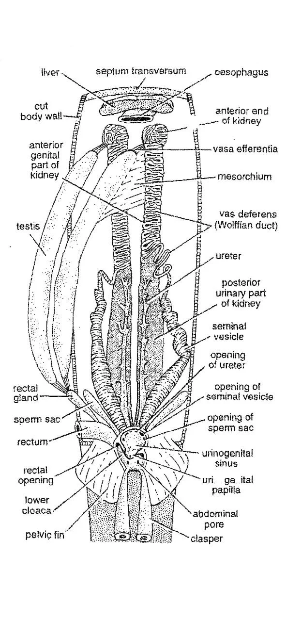
A. Male Excretory System
- A pair of kidneys are the main excretory organs of the male Scoliodon.
- The kidneys are mesonephric type.
- They are long, flattened, and ribbon-like.
- They are attached to the dorsal abdominal wall, above the peritoneum, one on either side of the median line.
- They extend nearly the whole length of the body cavity from the root of the liver in front up to the cloaca behind.
- The anterior part of the kidneys is non-excretory and they are genital in function. Hence they are called the epididymis.
- The posterior part is greatly developed and it is the main excretory part. The posterior part forms the functional adult kidney, called opisthonephros.
- Each kidney is formed by uriniferous tubules, Bowman’s capsule, and glomeruli.
- The tubules have a special urea-absorbing segment in them.
- Posteriorly, both the ureters open into a urinogenital sinus.
- The urinogenital sinus opens into the cloaca.
B. Male Reproductive System
- A pair of very large and elongated testes are the principal reproductive organs of male Scoliodon.
- They are attached to the mid-dorsal abdominal body wall by a double fold of peritoneum called mesorchium.
- From each testis, several very fine tubules are given off, called the vasa efferentia.
- The vasa efferentia run in the mesorchium to the anterior end of a large Wolffian or mesonephric duct now called the vas deferens.
- Spermatozoa developed from germ cells in seminiferous tubules of the testis by the process called spermatogenesis.
- The vas deferens forms a very large and extremely coiled duct that produces fluid for the nourishment of the spermatozoa.
- Posteriorly the vas deferens expands to form the seminal vesicle which is a wide straight tube.
- The seminal vesicles of both the sides open behind independently into the urinogenital sinus.
- The urinogenital sinus later opens into the cloaca.
- Claspers and siphon are the accessory parts of the male reproductive system.
- Claspers are present on the pelvic fins of male dogfish.
- Siphon is a muscular sac beneath skin of posterior ventral pelvic region.
- During copulation, sea water is drawn into siphon and forced down the clasper groove flushing spermatozoa accumulated there into the female cloaca.
Female Urinogenital System of Scoliodon
Diagram
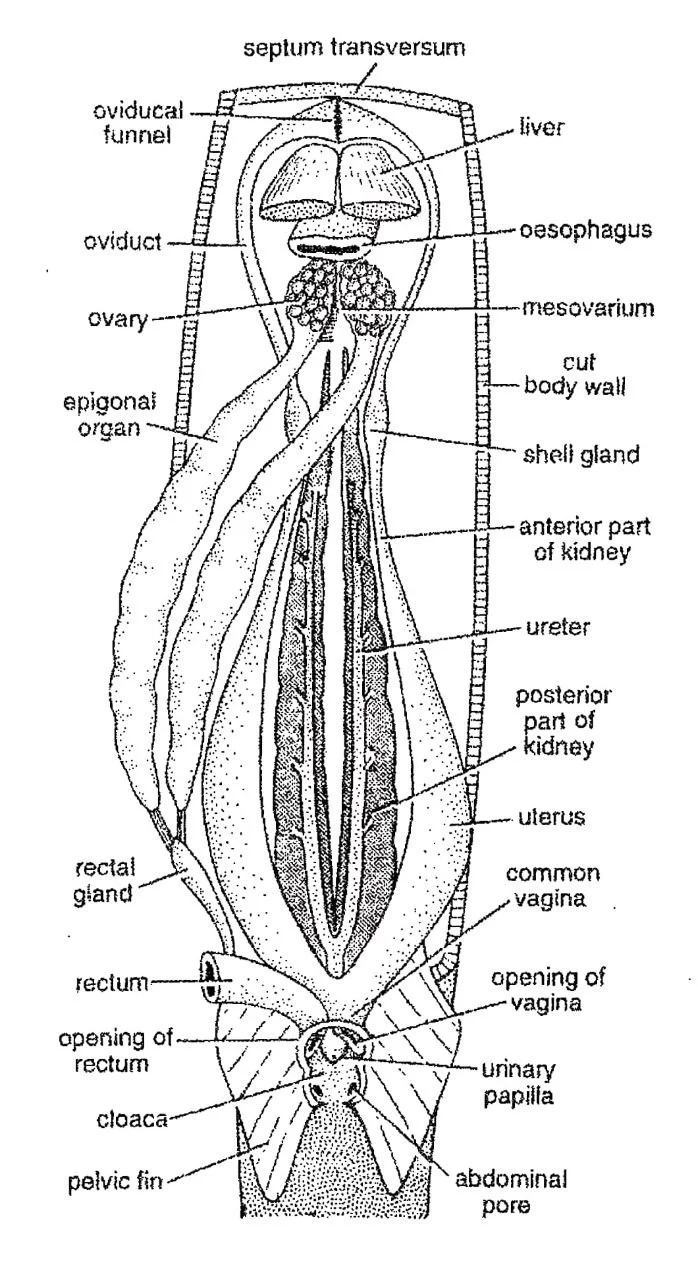
A. Female Excretory System
- Two kidneys are the main excretory organs.
- The kidneys are mesonephric type.
- The anterior portions of the kidneys are non-excretory.
- The posterior portions of the kidneys are the main excretory parts.
- In the female urinogenital system, there is no connection between the kidneys and genital organs.
- Therefore, the anterior part of the kidney is extremely reduced.
- The uriniferous tubules of the posterior functional part of the kidney open into a long thin-walled duct, the ureter.
- Unlike male, the two ureters of female dogfish unite into a common median ureter opening behind into the large median urinary sinus.
- The sinus later opens into the cloaca.
B. Female Reproductive System
- Female genital organs are one pair of small ovaries.
- They are attached one on either side mid-dorsally to the anterior abdominal wall by a fold of peritoneum, the mesovarium.
- Their form and size vary with the age.
- A pair of long tubular epigonal organs extend between ovaries.
- Mature ova are shed into the abdominal cavity from where they enter the oviducts.
- The two oviducts or Mullerian ducts are large tubes, extending the whole length of the body cavity.
- The two oviducts remain united both posteriorly and anteriorly.
- Anteriorly two oviducts unite mid-ventrally, below oesophagus to open into coelom by a single longitudinal slit, the ostium or oviducal funnel.
- Each oviduct bears a shell gland that secretes a thin membrane over the descending eggs.
- Posteriorly, two oviducts unite to form the vagina which opens into the cloaca.
——–THE END———
Read More:
- External Morphology of Scoliodon with Diagram | Dog fish
- Nervous System of Scoliodon | Shark | Diagram
- Reproduction of Scoliodon | Dog Fish
- Structure, Development & Homology of Placoid Scales | Shark | Scoliodon
- Respiratory System of Scoliodon | Dog Fish | Diagram
- Digestive System of Scoliodon with Diagram | Dog Fish | Note
- Sense Organs of Scoliodon | Diagram
- General Characters of All Classes of Vertebrates.
Reference:

Md Ekarm Hossain Bhuiyan is a dedicated zoology graduate with a profound passion for the study of animal life. He completed his primary and secondary education at Ispahani Public School and College, renowned for its commitment to academic excellence. He then pursued his secondary education at Government Science College. After that he achieved graduation at Department of Zoology, Jagannath University. His educational background and enthusiasm for zoology position him to make meaningful contributions to the field of biological sciences in Bangladesh.