The main receptor or sense organs of Scoliodon include (i) olfactory organs, (ii) eyes, (iii) ears, (iv) neuromasts or lateral line organs, and (v) ampullae of Lorenzini.
1. Olfactory Organs
- A pair of nasal or olfactory sacs are the main olfactory organs.
- They are characteristically correlated with a highly developed sense of smell for the perception of chemical substances dissolved in water.
- They are situated ventrally in the snout one on either side.
- Each olfactory sac is large and oval pouch.
- They are ectodermal blind pouch covered by a thin membrane.
- They are located inside the thin cartilaginous olfactory capsule of the skull.
- Each olfactory sac contains receptor and supporting cells.
- Sensory hairs project from the free ends of receptor cells.
- The basal ends of receptor cells are continued into fibres of olfactory nerve going to the olfactory lobe of the brain.
- Each olfactory sac opens to the outside ventro-laterally by the external nares.
2. Eyes
- They are also called photoreceptor organs.
- Scoliodon has a pair of large eyes.
- The eyes are located inside the orbits, one on either side of the head.
A. Eye Muscle
-
-
- The eyeball is elliptical in shape.
- The eyeball remains attached to the inner wall of the orbit by 6 eye muscles and a cartilaginous stalk called optic pedicle.
- There are two groups of eye muscles. They are oblique muscles and recti muscles.
- There are 2 oblique muscles and they are 1. inferior oblique muscle and 2. superior oblique muscle.
- The inferior oblique muscle is attached to the antero-ventral side of the eyeball.
- The superior oblique muscle is attached to the antero-dorsal side of the eyeball.
- The action of oblique muscles twists the eyeball.
- There are 4 recti muscles and they are 1. inferior rectus muscle, 2. superior rectus muscle, 3. anterior rectus muscle, and 4. posterior rectus muscle.
- The inferior rectus muscle is attached to the postero-ventral side of the eyeball.
- The superior rectus muscle is attached to the postero-dorsal side of the eyeball.
- The inferior and superior rectus muscles serve to rotate the eyeball in the vertical plane.
- The anterior (or internal) and posterior (or external) rectus muscles are inserted anteriorly and posteriorly.
- The anterior and posterior rectus muscles serve to rotate the eyeball in the horizontal plane.
-
B. Internal Structure
-
-
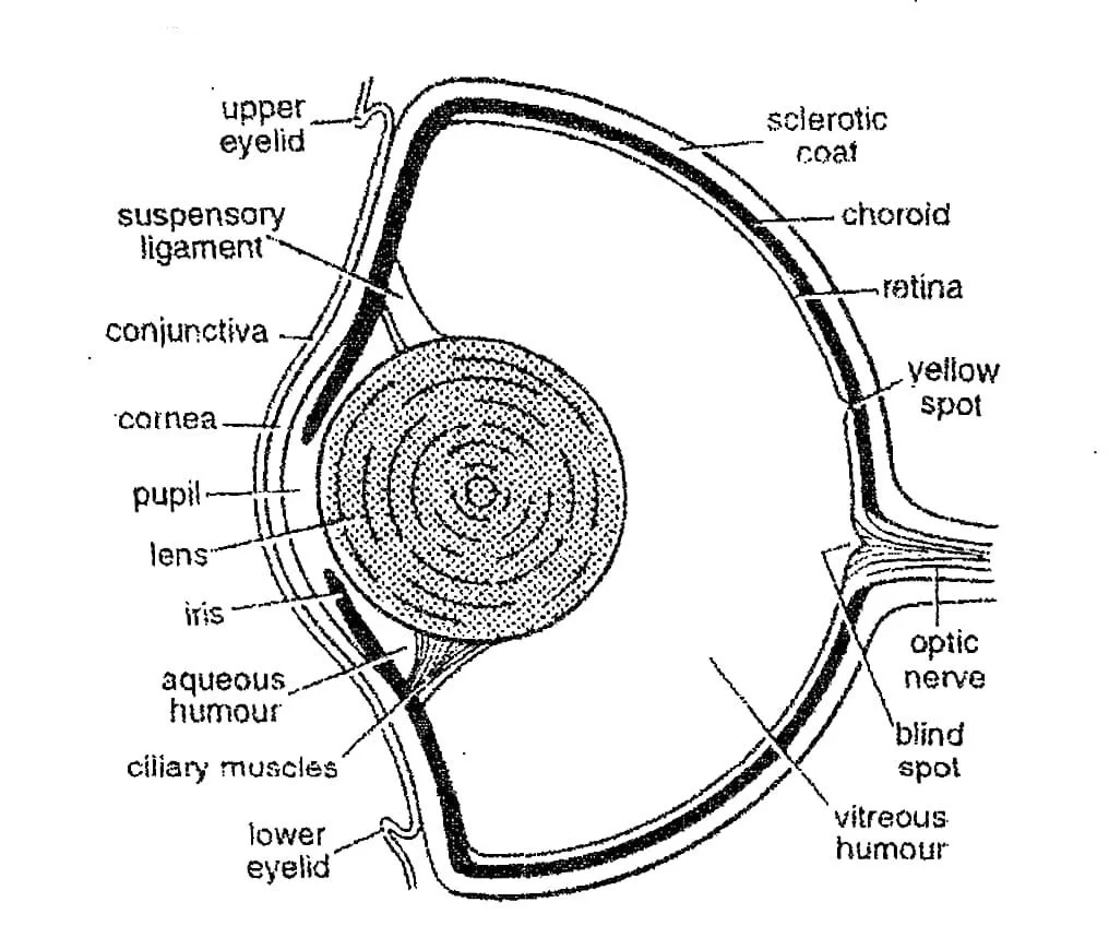
- The outermost layer of the eyeball is the sclerotic layer.
- The sclerotic layer is divided into two parts. The anterior part is the cornea and the posterior part is the white of the eye-ball.
- Cornea is transparent and it permits the light rays to enter into the eye. It is covered with thin and transparent conjunctiva.
- Choroid layer is located underneath the sclerotic layer.
- Choroid is a richly vascularized and pigmented layer.
- At the anterior end of the choroid, just behind the cornea, the choroid forms a pigmented circular disc called the iris.
- The iris has an aperture at the center named pupil. The whole eyeball is lightproof except the pupil.
- A large crystalline lens lies immediately behind the pupil. The pupil can not contract and dilate.
- Suspensory ligaments are located just behind the iris and they keep the lens in position.
- The innermost layer of the eyeball is the retinal layer.
- This is the only light-sensitive part of the eyeball.
- The retina consists mainly of elongated receptor cells, or rods, which are connected with the fibres of the optic nerve. Scoliodon has no cone cells.
- A transparent saline fluid, the aqueous humour, fills the small chamber in front of the lens.
- A jelly-like mass, the vitreous humour, fills the very large chamber behind the lens.
-
C. Mechanism of Sight/Vision
-
-
- Eyes in sharks are large but far separated, so binocular vision is not possible.
- The power of accommodation is poor because of the non-contractile pupils and little change in the shape of the lens.
- However, the lens can be shifted forward to catch more light.
- Since the retina is without cones, the fish is colour-blind and the eye cannot discriminate minute details.
- But the eye is adapted for near vision in dim light and fish can see its prey.
- Light rays pass through the cornea, pupil, and lens and reach the retina.
- The image formed by the retina is transmitted to the brain with the help of optic nerves.
-
3. Internal Ear
- In Scoliodon, there are no external or middle ears.
- Only internal ear is present called the membranous labyrinth.
- It is a delicate membranous ectodermal sac.
- It is found embedded in the cartilaginous olfactory capsule, one on either postero-lateral side of the cranium.
- The membranous labyrinth is innervated by the auditory nerve.
- Internal ears or membranous labyrinths of Scoliodon are also called stato-acoustic organs because of their two important functions. The functions are balancing and hearing.
4. Neuromast or lateral Line System
- It is a system of sense organs concerned with life in water.
- In Scoliodon it includes (a) lateral lines, (b) neuromast organs, and (c) pit organs.
a) Lateral Lines
-
-
- A lateral line runs along either lateral side of trunk and tail.
- It contains a mucus-filled ectodermal canal.
- It opens to the surface by minute pores.
-
b) Neuromast Organs
-
-
- These are small groups of receptor and supporting ectodermal cells located in the lateral line canals.
- Receptor or sensory cells bear tiny stiff sensory hairs.
- They are supplied with nerve fibres of facial, glossopharyngeal or vagus nerves.
- Neuromast organs are rheoreceptors or current receptors.
- They can perceive very low-frequency vibrations.
-
c) Pit Organs
-
-
- Small ectodermal pits are found scattered on the dorsal and lateral surfaces of head of Scoliodon.
- Each pit organ consists of sensory hair cells with supporting cells.
- They are innervated by nerve fibres of the VII cranial nerve.
-
5. Ampullae of Lorenzini
- These are situated in grape-like bunches in the snout of Scoliodon.
- They open on the dorsal and ventral surfaces of the head by their minute apertures.
- Each ampulla or ampullary sac is supplied with a nerve fibre from the branches of the VII cranial nerve.
===== THE END =====
Read More:
- Nervous System of Scoliodon | Shark | Diagram
- Reproduction of Scoliodon | Dog Fish
- Structure, Development & Homology of Placoid Scales | Shark | Scoliodon
- External Morphology of Scoliodon with Diagram | Dog fish
- Urinogenital System of Scoliodon | Diagram | Note
- Respiratory System of Scoliodon | Dog Fish | Diagram
- Digestive System of Scoliodon with Diagram | Dog Fish | Note
- General Characters of All Classes of Vertebrates.

Md Ekarm Hossain Bhuiyan is a dedicated zoology graduate with a profound passion for the study of animal life. He completed his primary and secondary education at Ispahani Public School and College, renowned for its commitment to academic excellence. He then pursued his secondary education at Government Science College. After that he achieved graduation at Department of Zoology, Jagannath University. His educational background and enthusiasm for zoology position him to make meaningful contributions to the field of biological sciences in Bangladesh.