The nervous system of Scoliodon consists of three parts :
1. Central nervous system.
2. Peripheral nervous system.
3. Autonomic nervous system.
1. Central Nervous System of Scoliodon
- It includes the brain and spinal cord of the Scoliodon.
A. The Brain of Scoliodon
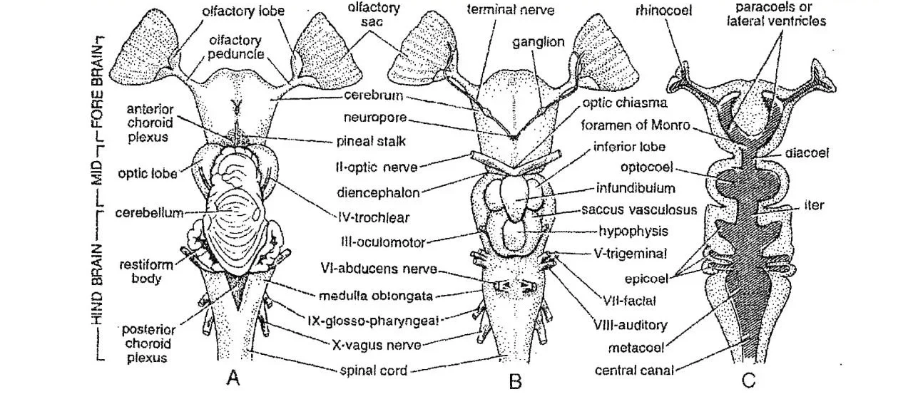
-
-
- The brain of Scoliodon lies enclosed within the chondrocranium.
- It is covered with meninges.
- It is made of the same three basic parts forebrain, midbrain, and hindbrain.
-
I) Forebrain
-
-
- It is the anterior part of the brain.
- It is subdivided into two parts and they are cerebrum and diencephalon.
-
a) Cerebrum
-
-
- It is also known as the cerebral hemisphere.
- It is the anteriormost part of the brain as well as the forebrain.
- It is undivided and has no mid-dorsal groove indicating the right and left cerebral hemispheres of higher vertebrate animals.
- From either antero-lateral side of the cerebrum arises an olfactory lobe.
- The midventral portion of the cerebrum has a small aperture called the neuropore.
- A terminal nerve comes out of each neuropore to innervate the mucous membrane of the olfactory sacs.
-
b) Diencephalon
-
-
- Diencephalon is located posterior to the cerebrum.
- It is completely hidden by the cerebellum.
- The roof of the diencephalon is called the anterior choroid plexus.
- A pineal body or epiphysis of unknown function is present at the mid-dorsal region of the diencephalon.
- Ventrally, at the anterior margin of the diencephalon, lies the optic chiasma.
- The optic chiasma is formed by the crossing of the two optic nerves.
- Just behind the chiasma, the floor gives off the infundibulum.
-
II) Midbrain
-
-
- It is the middle portion of the brain.
- It consists of a pair of large and rounded option lobes or corpora bigemina.
- Optic lobes are mainly responsible for the sense of sight.
- The oculomotor and trochlear cranial nerves arise from the midbrain.
-
III) Hindbrain
-
-
- It is the posterior portion of the brain.
- It comprises two parts, cerebellum and medulla oblongata.
-
a) Cerebellum
-
-
- It is rhomboidal in shape.
- The dorsal surface of the cerebellum shows irregular folds
- It is divided into three lobes by two transverse furrows.
- Sometimes, a median longitudinal furrow may also divide the cerebellum into right and left halves.
- It is the center of co-ordination of body movement.
-
b) Medulla Oblongata
-
-
- It is the posteriormost part of the brain.
- It is triangular in shape.
- It continues posteriorly into the spinal cord.
- V, VI, VII, VII, IX and X cranial nerves arise from the medulla oblongata.
-
B. Spinal Cord of Scoliodon
-
-
- The spinal cord extends from the medulla oblongata almost to the end of the tail.
- It is protected by the neural canal of vertebrae.
- In structure, it resembles that of higher vertebrates in having dorsal and ventral fissures, central canal, and inner grey and outer white matter.
-
2. Peripheral Nervous System of Scoliodon
- It includes cranial nerves and spinal nerves of the Scoliodon.
A. Cranial Nerves of Scoliodon
-
-
- The brain gives off 10 pairs of cranial nerves.
- They are identified by their serial Roman numbers as well as names.
-
All ten pairs of cranial nerves of Scoliodon are given below:-
| Serial | Name | Type | Origin | Distribution |
| I | Olfactory nerves | Sensory | Olfactory lobe | Olfactory sac. |
| II | Optic nerves | Sensory | Optic thalamus or ventral side of the diencephalon | The retina of the eye. |
| III | Oculomotor nerves | Motor | Ventral surface of the midbrain | Recti and oblique muscles of eye. |
| IV | Trochlear or Pathetic nerves | Motor | Dorso-laterally from midbrain | Oblique muscles of the eye. |
| V | Trigeminal nerves | Mixed | Antero-lateral side of the medulla oblongata | Snout, upper & lower jaws, tongue, etc. |
| VI | Abducens nerves | Motor | Midventrally from
medulla oblongata |
Recti muscles of the eye. |
| VII | Facial nerves | Mixed | Medulla oblongata | Ampullae of Lorenzini., snout, buccal cavity, throat, mandible, etc. |
| VIII | Auditory nerves | Sensory | Medulla oblongata | membranous
labyrinth(internal ear). |
| IX | Glossopharyngeal
nerves |
Mixed | Ventrolaterally from the medulla oblongata | Pharynx |
| X | Vagus nerves | Mixed | Posterior lateral side of the medulla oblongata | Heart, digestive tract, neuromast organs, pharynx, etc. |
B. Spinal Nerves of Scoliodon
-
-
- Several pairs of spinal nerves arise from spinal cord.
- Each nerve, originates by two roots, a sensory dorsal root and a motor ventral root.
- Each spinal nerve divides into three branches; ramus dorsalis, ramus ventralis and ramus communicans.
-
3. Autonomic Nervous System of Scoliodon
- It includes a series of paired and irregularly arranged ganglia in the dorsal wall of posterior cardinal sinuses, and in dorsal parts of kidneys.
- The gastric ganglion is the largest ganglion.
- There is usually one ganglion in each segment.
———– THE END ———-
Read More:
- Sense Organs of Scoliodon | Diagram
- Reproduction of Scoliodon | Dog Fish
- Structure, Development & Homology of Placoid Scales | Shark | Scoliodon
- External Morphology of Scoliodon with Diagram | Dog fish
- Urinogenital System of Scoliodon | Diagram | Note
- Respiratory System of Scoliodon | Dog Fish | Diagram
- Digestive System of Scoliodon with Diagram | Dog Fish | Note
- General Characters of All Classes of Vertebrates.
Reference:

Md Ekarm Hossain Bhuiyan is a dedicated zoology graduate with a profound passion for the study of animal life. He completed his primary and secondary education at Ispahani Public School and College, renowned for its commitment to academic excellence. He then pursued his secondary education at Government Science College. After that he achieved graduation at Department of Zoology, Jagannath University. His educational background and enthusiasm for zoology position him to make meaningful contributions to the field of biological sciences in Bangladesh.