In this article about the Scoliodon, we will learn about the digestive system of Scoliodon with a diagram. Scoliodon is a cartilaginous fish that belongs to the class Chondrichthyes.
What is Digestive System?
The digestive system is a physiological system by which an animal turns complex food into absorbable simple food in order to keep functioning the body.
Digestive System of Scoliodon
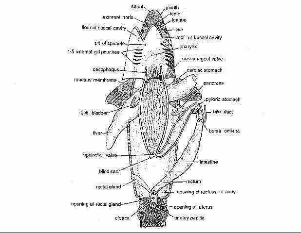
fig: Digestive system of Scoliodon.
The entire digestive system of Scoliodon is divided into two major parts. They are 1. Alimentary canal, and 2. Digestive glands.
1. Alimentary Canal
- The alimentary canal starts from the mouth and ends at the anus.
- The digestive system of Scoliodon is complete because it has both mouth and anus.
- The alimentary canal is longer than the body of the dogfish.
- It consists of the mouth, buccal cavity, pharynx, oesophagus, stomach, intestine, and anus.
A. Mouth
-
-
- The alimentary canal starts from here.
- It is a wide crescentic opening.
- It is present on the ventral side of the head.
- It is bounded by upper and lower lips.
-
B. Buccal Cavity
-
-
- The mouth opens into the buccal cavity.
- It is spacious, dorso-ventrally flattened, and lined with mucous membrane.
- Jaws are present in the buccal cavity.
- Teeth are present but they are not attached to the jaws. Teeth are embedded in the skin.
- Teeth are homodont type, which means all the teeth are similar in shape.
- Teeth are sharply pointed and directed backward. They are arranged in several rows.
- Teeth can be replaced several times if lost or destroyed. That’s why their teeth are polyphyodont.
- The tongue is present on the floor of the buccal cavity.
- The tongue of Scoliodon is non-muscular and non-granular.
-
C. Pharynx
-
-
- The buccal cavity leads into the pharynx.
- It is lined by endoderm.
- On either side of the pharynx, there present internal openings of the spiracles and five branchial clefts.
- The mucous membrane of the pharyngeal wall bears numerous dermal denticles.
-
D. Oesophagus
-
-
- The pharynx leads into the oesophagus.
- It is a narrow and short tube.
- It has thick muscular wall.
- The mucous membrane of the oesophagus forms longitudinal folds.
-
E. Stomach
-
-
- The oesophagus opens into the stomach.
- It is present in the abdominal cavity of the Scoliodon.
- It is highly muscular and bent itself to form a “U” shaped structure.
- The stomach is divided into two parts and they are the cardiac stomach and the pyloric stomach.
- The proximal limb of the stomach is called the cardiac stomach and it is longer and wider than the distal limb.
- The distal limb of the stomach is called the pyloric stomach and it is shorter and narrower than the cardiac stomach.
- The oesophageal opening into the cardiac stomach is guarded by a sphincter called the oesophageal valve.
- At the junction of the cardiac and pyloric stomach, a small outgrowth is present, called the blind sac.
- The mucous lining of the cardiac stomach forms longitudinal folds and the mucous lining of the pyloric stomach is mostly smooth.
- At the end of the pyloric stomach, a muscular sphincter is present, called the pyloric valve.
- The opening of the pyloric stomach into a small chamber called the bursa entiana is guarded by the pyloric valve.
-
F. Intestine
-
-
- The bursa entiana is followed by the intestine.
- It is a straight and wide tube.
- The middle part of the intestine is as wide as the cardiac stomach.
- The anterior part of the intestine receives the bile and pancreatic ducts.
- In Scoliodon, the inner mucous lining of the intestine becomes folded anticlockwise into a longitudinal spiral or scroll of about two and a half turns. This is called the scroll valve or spiral valve.
-
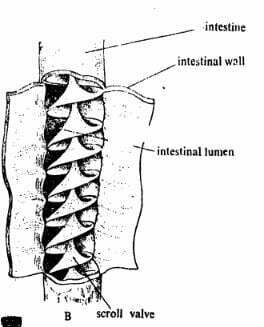
Fig: Scroll valve of Scoliodon
-
-
- The posterior part of the intestine is the rectum.
- It is a short and narrow tube where the faecal matter is stored.
- The rectum opens into the cloaca.
-
G. Anus
-
-
- The anus is present in the cloaca.
- The Scoliodon expels its faecal matter through this opening.
- The alimentary canal ends here.
-
2. Digestive Glands
Digestive glands are also an important part of the digestive system of Scoliodon. The digestive glands are described below-
A. Liver
-
-
- It is a bilobed gland which means it has two lobes.
- It is a yellowish and large gland.
- The two lobes are attached at the anterior and free at the posterior portion.
- A thin-walled “V” shaped gall bladder is present at the anterior portion of the right lobe.
- In the gall bladder, bile is stored. The liver secretes the bile.
- A bile duct arises from the gall bladder and opens into the anterior part of the intestine.
- The bile duct also receives branches from the lobes of the liver.
-
B. Pancreas
-
-
- It is also a bilobed gland and whitish in color.
- The two lobes are the longer dorsal lobe and the smaller ventral lobe.
- A small pancreatic duct arises from the pancreas and opens into the anterior part of the intestine.
-
C. Caecal or rectal gland
-
-
- It is present on the dorsal side of the rectum.
- It is highly vascular and composed of lymphoid tissue but discharges a fluid in the intestinal lumen.
-
D. Spleen
-
-
- It is a large gland closely attached to the stomach.
- It is morphologically connected to the alimentary canal but it has no relation to the alimentary canal. It is functionally associated with the circulatory system.
-
3. Physiology of Digestion
In the digestive system of Scoliodon, the physiology of digestion of explained below-
- Scoliodon is a predatory and carnivorous fish.
- Its main food is other fishes. Sometimes it also eats crabs, lobsters, and worms.
- As the buccal cavity lacks salivary glands, no digestion occurs here.
- With the help of gastric juice in the stomach, food becomes digested.
- The gastric juice in the stomach contains pepsin and hydrochloric acid.
- The semi-digested food from the stomach is transferred into the intestine.
- Food becomes fully digested in the intestine with the help of bile and pancreatic juice.
- The complete absorption takes place inside the intestine.
——-THE END——
Read More:
- Respiratory System of Scoliodon | Dog Fish | Diagram
- Urinogenital System of Scoliodon | Diagram | Note
- External Morphology of Scoliodon with Diagram | Dog fish
- Nervous System of Scoliodon | Shark | Diagram
- Reproduction of Scoliodon | Dog Fish
- Structure, Development & Homology of Placoid Scales | Shark | Scoliodon
- Sense Organs of Scoliodon | Diagram
- General Characters of All Classes of Vertebrates.
References:

Md Ekarm Hossain Bhuiyan is a dedicated zoology graduate with a profound passion for the study of animal life. He completed his primary and secondary education at Ispahani Public School and College, renowned for its commitment to academic excellence. He then pursued his secondary education at Government Science College. After that he achieved graduation at Department of Zoology, Jagannath University. His educational background and enthusiasm for zoology position him to make meaningful contributions to the field of biological sciences in Bangladesh.