In this article, we will learn about the digestive system of frog Rana tigrina. Rana tigrina or Hoplobatrachus tigerinus is the scientific name of the Indian Bull Frog. Previously it was named Rana tigrina but now the updated and new scientific name of the Indian Bull Frog is Hoplobatrachus tigerinus. It is a large species of frog in South and Southeast Asia.
Systematic Position of Indian Bull Frog
Kingdom: Animalia
Phylum: Chordata
Subphylum: Vertebrata
Class: Amphibia
Order: Anura
Family: Dicroglossidae
Genus: Hoplobatrachus
Species: Hoplobatrachus tigerinus
Digestive System of Frog (Rana tigrina or Hoplobatrachus tigerinus)
Digestion is a physiological system by which the complex food turns into simple food to absorb nutrients to keep the body functioning and healthy. The digestive system is a system of organs by which digestion occurs. The digestive system of frog is divided into two parts. They are:
1. Digestive Tract / Alimentary Canal, and
2. Digestive Glands.

fig: Digestive system of frog
1. Alimentary Canal
- The alimentary canal of the frog is complete.
- It is a long coiled tube.
- It starts from the mouth and ends at the cloacal aperture.
- The alimentary canal consists of –
- Mouth
- Buccal cavity
- Pharynx
- Oesophagus
- Stomach
- Small Intestine
- Large Intestine
- Cloaca
A. Mouth
-
- The alimentary canal of frog starts from the mouth.
- The mouth is the transverse and largest aperture of the body.
- It is a wide transverse slit-like aperture.
- The mouth extends from one side of the snout to another side of the snout.
- It is bounded by two bony jaws.
- The upper jaw is fixed and the lower jaw can move up and down.
- Lips cover the jaws and the lips are immovable.
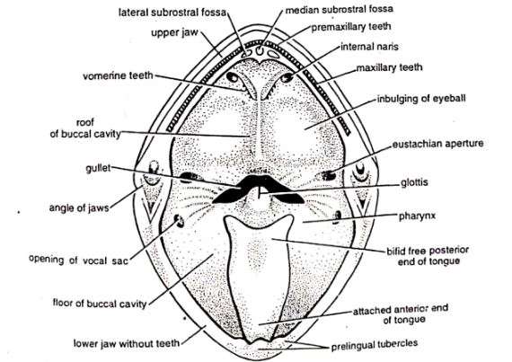
fig: Bucco-Pharyngeal cavity of a frog.
B. Buccal cavity
-
- The mouth leads into the buccal cavity.
- It is large, wide, and shallow.
- It has ciliated columnar epithelial lining.
- The buccal cavity has mucous glands which secret mucous to lubricate the food.
- There are no salivary glands present in frogs. So no saliva secrets and no digestion occurs in the buccal cavity.
- The buccal cavity has teeth, tongue, internal nostrils, and bulging of orbits.
Teeth
-
-
-
- The premaxillae and maxillae bones of the upper jaw contain teeth.
- Teeth occur in a row and on either side of the premaxillae and maxillae bones.
- Teeth are small, conical, and backwardly pointed.
- The lower jaw has no teeth.

- fig: teeth of frog
-
- Two small bones are also present on the roof of the mouth called vomers and they bear vomerine teeth.
- The frogs can not chew with their teeth. The teeth are just for holding the prey and preventing the prey from slipping out.
- The teeth of the frogs are homodont and acrodont. Homodont means all the teeth are similar and acrodont means the teeth are not set in a socket.
- Teeth are attached to the jaw by a broad base.
- The free part of a tooth is called the crown. It is made up of dentine. The tip of the crown is covered with enamel which is a very hard substance.
- The tooth contains a central pulp cavity open at the side and filled with a soft nourishing pulp.
- The teeth of frogs are replaced several times when they are lost and that’s why frogs are polyphyodont.
-
-
Tongue
-
-
-
- The tongue is present on the floor of the mouth cavity.
- It is large, muscular, sticky, and protrusible.
- Its anterior end is attached to the inner border of the lower jaw and the posterior end is free.
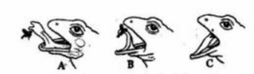
- fig: Preying mechanism of frog with tongue
-
- The posterior end of the tongue can be flicked out and retracted immediately after catching the prey with its slimy surface.
- The protrusion of the tongue is brought about by the change of pressure in the large sublingual lymph sac.
-
-
Internal Nostrils
-
-
-
- Located on the roof of the buccal cavity.
- Just in front of the vomerine teeth, a pair of small openings present which are called internal nostrils or internal nares.
- These internal nostrils connect the nasal cavity and the buccal cavity.
- These openings are the part of frog’s respiration.
-
-
Bulgings of Orbits
-
-
-
- Bulgings of orbits or eyeballs are present on the roof of the buccal cavity and behind the vomerine teeth.
- These are large and oval in shape.
- While swallowing the food, the frog depresses the eyes inwards and pushes the food inside the pharynx.
-
-
C. Pharynx
-
- The buccal cavity leads into the pharynx.
- It is short in size.
- As the pharynx is short in size, sometimes both the buccal cavity and the pharynx are described as bucco-pharyngeal cavity.
- Various apertures open into the pharynx.
- There is a longitudinal slit-like aperture present on the floor of the pharynx named glottis.
- The glottis leads to the laryngo-tracheal chamber.
- On the roof on either lateral side, there is a wide eustachian aperture.
- The eustachian aperture opens into the middle ear.
- In male frog, near the angle of two jaws, on the floor of the pharynx on either side, is present the small opening of a vocal sac.
- The pharynx leads into oesophagus through a wide opening named the gullet.
D. Oesophagus
-
- The pharynx leads into the oesophagus.
- It is a short, wide and muscular tube.
- It is highly distensible.
- Its mucous epithelial lining contains some mucous glands.
- Its mucous epithelial lining is folded longitudinally and these foldings allow its expansion during the passage of food into the stomach.
- The glandular lining of the oesophagus secretes digestive fluid.
- Oesophagus enlarges to merge with the stomach in the peritoneal cavity.
E. Stomach
-
- The oesophagus leads into the stomach.
- It lies on the left side of the body cavity.
- The stomach is attached to the dorsal bodywall by a mesentery called mesogaster.
- It is large and broad and almost 4 cm in length.
- It is a slightly curved sac-like tube and has thick muscular walls.
- The stomach is divided into two parts.
- The anterior broader part of the stomach is called the cardiac stomach and the posterior narrower part is called the pyloric stomach.
- The inner surface of the stomach has several longitudinal folds which allow distension when the food is received.
- Inside the stomach, gastric glands secret the enzyme pepsinogen and oxyntic glands secret hydrochloric acid.
- The posterior part of the stomach, the pyloric stomach, opens into the small intestine.
- The opening of the pyloric stomach into the small intestine is guarded by a sphincter muscle called the pyloric valve.
- The stomach works as the storage as well as the digestion of food.
F. Small Intestine
-
- The stomach opens into the small intestine.
- It is a long, coiled, and narrow tube.
- It is about 30 cm long.
- The small intestine is attached mid-dorsally to the body wall by mesenteries.
- The mucosal lining of the small intestine consists of two types of cells besides intestinal glands. They are large goblet cells and small absorbing cells.
- Goblet cell contains oval vacuoles and granular substances which possibly produces mucous.
- The nucleus of the goblet cell is situated near the base of the cell.
- The small intestine is made up of two parts and they are 1. small anterior duodenum and 2. large posterior ileum.
Duodenum
-
-
-
- It is the anterior part of the small intestine.
- It runs ahead parallel to the stomach.
- The stomach and the duodenum form a “U” shape.
- It receives the hepatopancreatic duct.
- The hepatopancreatic duct comes from the liver and the pancreas.
- With the help of the hepatopancreatic duct, bile and pancreatic juice come to the small intestine.
- The internal mucosal lining forms low transverse folds.
-
-
Ileum
-
-
-
- It is the posterior part of the small intestine.
- It is much longer than the duodenum.
- Also, it is the longest part of the alimentary canal.
- Before joining to the rectum posteriorly, it forms several folds.
- The internal mucosal lining forms many longitudinal folds.
- There are no true villi and definite glands in the ileum.
- Both digestions of food and absorption of digested food occur in the small intestine.
-
-
G. Large Intestine
-
- The large intestine is also known as the rectum.
- It is short in size but a wide tube.
- It is approximately 4 cm in length.
- It runs straight behind to open into the cloaca.
- The large intestine is guarded by the anal sphincter before opening into the cloaca.
- The inner lining of the rectum forms numerous longitudinal folds to re-absorb the water.
- The function of the large intestine is to re-absorb the water and prepare and store the faeces.
H. Cloaca
-
- Alimentary canal ends at the cloaca.
- It is a small sac-like part.
- Anus and urinogenital apertures are open here.
- The cloaca opens to the outside by cloacal aperture which is located at the hind end of the body, between the two hind limbs.
2. Digestive Glands
Besides gastric glands and intestinal glands, two large digestive glands are associated with the digestive system of frog and they are:
A. Liver, and
B. Pancreas
A. Liver
-
- The liver is the largest gland of the body.
- In fact, the liver is the largest gland of all vertebrates.
- The colour of the liver is reddish-brown.
- It is located close to the heart and lungs.
- The liver is divided into 3 lobes and they are right, left, and median.
- The innumerable polygonal cells of the liver secret a greenish alkaline fluid called the bile.
- Bile is stored in the gall bladder.
- The gall bladder is a thin-walled sac that lies between the lobes of the liver.
- Cystic ducts from the gall bladder and hepatic ducts from the liver lobes combine and form a common bile duct.
- The common bile duct then joins with the pancreatic duct and forms a hepatopancreatic duct. And this hepatopancreatic duct then opens into the duodenum.
- Bile has no digestive ferments; it only emulsifies the fats and that’s why the liver is not truly a digestive gland.
B. Pancreas
-
- It is located between the stomach and duodenum.
- It is a branched, irregular, flattened, and yellow-coloured gland.
- It works as both endocrine and exocrine.
- The endocrine of the pancreas is formed by islets of Langerhans which produce the hormone Insulin.
- Insulin is concerned with sugar metabolism.
- The exocrine of the pancreas secrets pancreatic juice.
- Pancreatic juice contains several digestive enzymes.
- The pancreas has no independent ducts and it serves its pancreatic juice to the duodenum through the hepatopancreatic duct.
Mechanism of Digestion
- Frog is strictly carnivorous and it feeds on insects, worms, crustaceans, small fishes, and even small frogs and tadpoles.
- They prey on their food with their tongue and swallow the whole food without mastication.
- Food passes into the stomach through the mouth, buccal cavity, pharynx, and oesophagus.
- Food is pushed down the oesophagus by a wave of contraction of its muscular wall called peristalsis.
- Food stays in the stomach for 2-3 hours.
- Hydrochloric acid and pepsinogen are secreted by the gastric glands. Pepsinogen catalyzes the proteins into peptones and proteases. Hydrochloric acid kills bacteria and fungi and softens the food particles.
- Then the food passes into the small intestine.
- In the intestine bile, pancreatic juice and intestinal juice mix up with the food.
- Digestion is completed inside the small intestine.
- The absorption occurs in the small and large intestine.
- Undigested food particles then move to the rectum and are eventually expelled out through the cloacal aperture.
- The digestive system of frog is complete as it starts from the mouth and ends at the cloaca.
———THE END———-
Read More:
- External Morphology of Frog | Diagram
- Blood Circulatory System of Frog | Diagram
- Urinogenital System of Frog | Diagram
- Nervous System and Sense Organs of Frog | Diagram
- Respiratory System of Frog | Diagram
- Hoplobatrachus tigerinus (Previously Rana tigrina) | Indian Bull Frog
- General Characters of All Classes of Vertebrates.
Reference:
2. Frog wiki

Md Ekarm Hossain Bhuiyan is a dedicated zoology graduate with a profound passion for the study of animal life. He completed his primary and secondary education at Ispahani Public School and College, renowned for its commitment to academic excellence. He then pursued his secondary education at Government Science College. After that he achieved graduation at Department of Zoology, Jagannath University. His educational background and enthusiasm for zoology position him to make meaningful contributions to the field of biological sciences in Bangladesh.