Blood Circulatory System of Frog
- The blood circulatory system of frog is closed.
- The whole blood circulatory system of frog consists of blood, heart, arterial system, venous system.
Blood of Frog
- It is the chief circulatory fluid of the body.
- It contains blood plasma and blood corpuscles.
- It is a liquid connective tisse.
1. Blood Plasma
-
-
- It forms the two third part of the blood.
- Almost 90% of the blood plasma is water.
- It contains mineral salts, absorbed food (sugars, proteins), excretory wastes, hormones and other soluble substances.
-
2. Blood Corpuscles
-
-
- There are three types of blood corpuscles and they are erythrocytes, leukocytes and thrombocytes.
- Erythrocytes or red blood corpuscles are oval, nucleated and flattened.
- Erythrocytes bear the respiratory pigment haemoglobin which carry oxygen to the tissues.
- Leukocytes or white blood corpuscles are colorless, nucleated and amoeboid cells.
- Leukocytes are phagocytic, ingesting bacteria and other foreign particles that arrive in blood.
- Thrombocytes or blood platelets play an important role in blood coagulation.
-
Heart of Frog
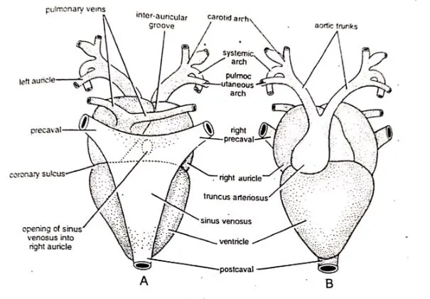
Fig : Heart of frog; A – Dorsal view; B – Ventral view.
External Features
- It lies mid-ventrally inside the anterior trunk region.
- It is protected by the pectoral girdle.
- It is reddish in color.
- It is somewhat conical or triangular in shape.
- Its broad base is directed anteriorly and the narrow apex is directed posteriorly.
a) Pericardium
-
-
- The heart is enclosed by a sac, called the pericardium.
- It is a thin, transparent, and two-layered sac.
- The outer wall of the pericardium is called the parietal pericardium and the inner wall is called the visceral pericardium.
- Between the two pericardial layers, there is a narrow cavity that contains a fluid called the Pericardial fluid.
- The pericardial fluid protects the heart from friction or mechanical shocks.
-
b) Chambers of Heart
-
-
- Frog’s heart is three chambered; two auricles and a ventricle.
- The two auricles are externally demarcated by the inter-auricular groove.
- The two auricles are clearly marked off from the ventricle by the auriculo-ventricular groove.
- The heart of frog has two additional chambers and they are sinus venosus and truncus arteriosus.
- Sinus venosus is formed by the union of three large caval veins; two anterior precavals and one posterior postcaval.
- The truncus arteriosus bifurcates into two branches and each branch further divides into three arches and they are carotid, systematic and pulmocutaneous.
-
Internal Features
a) Auricles
-
-
- The two auricles are thin-walled.
- They are completely separated from each other by the inter-auricular septum.
- The right auricle is larger than the left.
- The sinus venosus opens into right auricle through the sinu-auricular aperture.
- Both auricles open into the ventricle through the auriculo-ventricular aperture which is guarded by the auriculo-ventricular valves.
-
b) Ventricle
-
-
- The ventricle has thick muscular and spongy wall.
- The cavity of ventricle is greatly reduced.
- The flaps of auriculo-ventricular valves are connected to the wall of ventricle by thread-like chordae tendineae.
-
c) Truncus Arteriosus
-
-
- The opening of ventricle into truncus arteriosus is guarded by three semilunar valves.
- The valves prevent the backflow of blood.
- The truncus arteriosus bifurcates into two branches and each branch further divides into three arches and they are carotid, systematic and pulmocutaneous.
-
Working of Heart
- Contraction of the heart is called systole and the relaxation of the heart is called diastole.
- Sinus venosus receives the deoxygenated blood from the venous system.
- Then the sinus venosus contracts. The deoxygenated blood from sinus venosus is forced to the right auricle.
- Meanwhile the oxygenated blood from the lungs is poured into the left auricle.
- Then the two auricles contract to force their blood into the ventricle.
Arterial System of Frog
- Arteries carry blood away from the heart.
- The arterial system of frog begins with the truncus arteriosus.
- The truncus arteriosus divides into right and left branches. Both branches later subdivides into three major aortic arches and they are common carotid arch, systemic arch and pulmocutaneous arch.
- The common carotid arch divides into two branches and they are external carotid and internal carotid.
- The common carotid arch supplies blood to the head.
- The systemic arch is the longest of the three arches.
- The two systemic arches gives off three arteries and they are oesophageal, occipito-vertebral and subclavian arteries.
- The both systemic arches later join with each other behind the heart to form the dorsal aorta.
- The dorsal aorta gives off five types arteries and they are coeliaco-mesenteric, gonadial, renal, posterior mesenteric and common iliac arteries.
- The pulmocutaneous arches divide into two arteries and they are pulmonary and cutaneos artery.
- The pulmonary artery supplies blood to the lungs and the cutaneous artery supplies blood to the skin.
Venous System of Frog
- The venous system collects blood and returns to the heart.
- The venous system has four parts and they are pulmonary veins, caval veins, renal portal veins, and hepatic portal veins.
- Oxygenated blood from the lungs are collected by the two pulmonary veins.
- There are three caval veins. Of them two are anterior vena cava or precavals and one is posterior vena cava or postcaval.
- All the caval veins open into the sinus venosus.
- Each precaval is formed by the union of three veins and they are external jugular, innominate and subclavian veins.
- The postcaval receives renal veins, genital veins and hepatic veins before opening into the sinus venosus.
- Frog has two well-developed portal systems. They are the renal portal system and the hepatic portal system.
- The veins which carry blood to a capillary system in the kidneys constitute the renal portal system.
- A large hepatic portal vein is formed by the union of several branches from stomach, intestine, pancreas and spleen.
——————-THE END——————-
Read More:
- Respiratory System of Frog | Diagram
- External Morphology of Frog | Diagram
- Digestive system of Frog with Diagram
- Urinogenital System of Frog | Diagram
- Nervous System and Sense Organs of Frog | Diagram
- Hoplobatrachus tigerinus (Previously Rana tigrina) | Indian Bull Frog
- General Characters of All Classes of Vertebrates.
Reference:

Md Ekarm Hossain Bhuiyan is a dedicated zoology graduate with a profound passion for the study of animal life. He completed his primary and secondary education at Ispahani Public School and College, renowned for its commitment to academic excellence. He then pursued his secondary education at Government Science College. After that he achieved graduation at Department of Zoology, Jagannath University. His educational background and enthusiasm for zoology position him to make meaningful contributions to the field of biological sciences in Bangladesh.