What is the Placoid Scale?
Placoid scales, also known as dermal denticles, are a type of scale that is found on the skin of cartilaginous fishes such as sharks, rays, and skates. These scales have a flattened, plate-like shape and are composed of dentin, a hard, mineralized substance similar to the material found in our teeth, and enamel, a hard, mineralized substance that covers the dentin.
Structure of a Placoid Scale

- Placoid scales are characteristic of the skin of dogfishes, sharks, rays, etc.
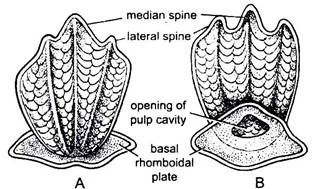
- A typical placoid scale consists of two parts: a basal plate and a spine.
- The basal plate is wide and rhomboidal and the spine is flat and trident.
- The spine arises from the center of the basal plate.
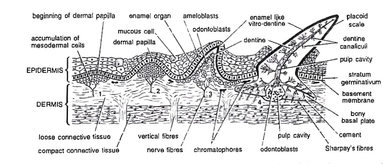
- The basal plate is embedded in the dermis and firmly held by Sharpey’s and other connective tissue fibers.
- The basal plate is formed by a bone-like loose trabecular calcified tissue called cement.
- The trident spine projecting out of the skin is backwardly directed.
- The spine is formed by one median and two lateral spines.
- In the vertical section, the spine consists chiefly of a hard substance called dentine with a central pulp cavity containing blood vessels, nerve endings, lymph channels and dentine-forming cells called odontoblasts.
- Through dentine ramify numerous very fine tubules or canaliculi containing long protoplasmic processes of odontoblasts.
- The spine is coated externally with a layer of still harder, shiny, enamel-like vitro-dentine.
Development of a Placoid Scale

- The first sign of a developing scale is an aggregation of mesodermal cells of the dermis, lying just beneath the epidermis and forming a dermal papilla.
- As the epidermis is pushed up, the outermost cells of the dermal papilla become odontoblasts (or scleroblasts) and deposit dentine between themselves and the epidermis.
- Meanwhile, the overlying epidermal cells of stratum germinativum or Malpighian layer, now called ameloblasts, form the so-called enamel organ which deposits vitrodentine over dentine.
- Cement (basal plate) is formed from the surrounding cells of the dermis.
- In a growing scale, the dermal papilla forms nutritive pulp occupying the pulp cavity.
- In a fully formed scale, the epidermis wears off around the spine which projects above the skin, while the basal plate remains embedded.
Homology of Placoid Scales

- The homology of placoid scales refers to the evolutionary origin and relationships of these scales to other structures in different species.
- It has been suggested that placoid scales are modified teeth, with a similar developmental origin and genetic basis.
- In fact, some scientists believe that placoid scales and teeth are homologous structures, based on similarities in their embryonic development and gene expression.
- Studies have shown that the genes involved in tooth development, such as BMP4 and SHH, are also expressed in the development of placoid scales.
- This suggests that both placoid scales and teeth structures may have a common evolutionary origin and may have diverged from a common ancestor.
- The placoid scales and teeth have essentially similar forms, structures, and embryonic development, indicating that they are homologous structures.
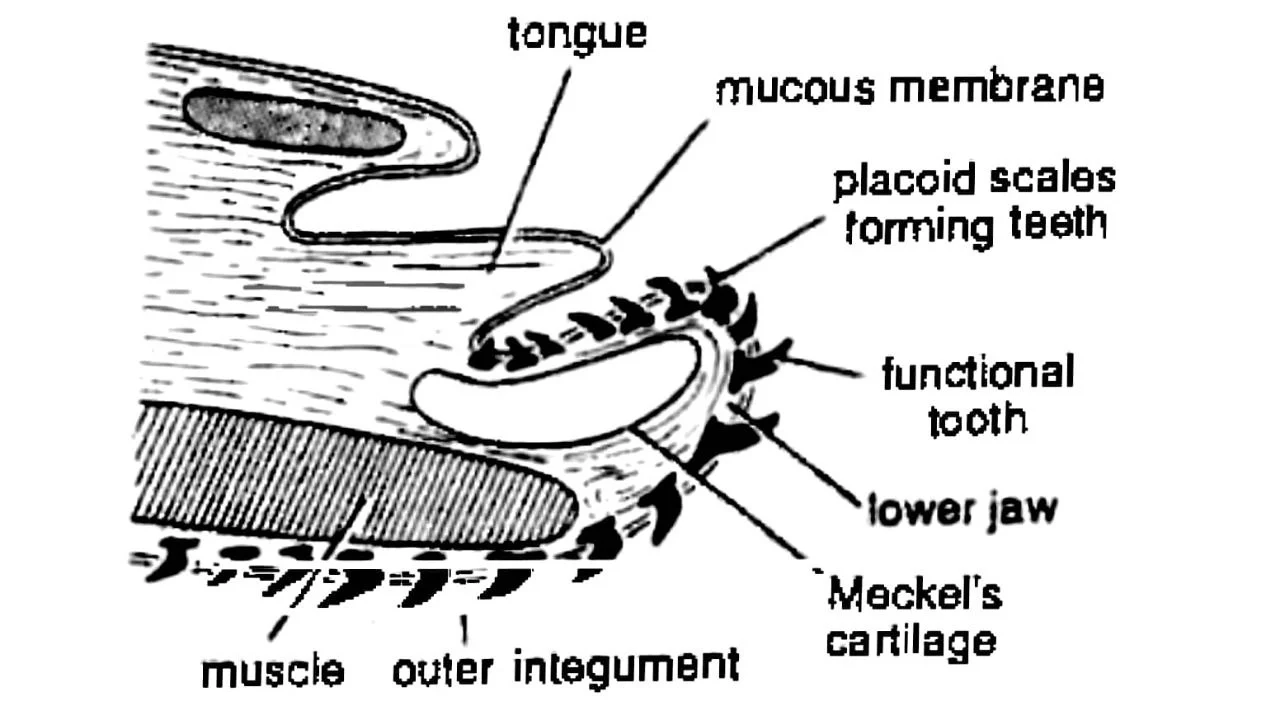
- The placoid scales become especially large to serve as teeth in the mouth.
- The scales are used for holding and tearing prey with their backwardly directed spines.
———— THE END ———–
Read More:
- Reproduction of Scoliodon | Dog Fish
- Nervous System of Scoliodon | Shark | Diagram
- Sense Organs of Scoliodon | Diagram
- External Morphology of Scoliodon with Diagram | Dog fish
- Urinogenital System of Scoliodon | Diagram | Note
- Respiratory System of Scoliodon | Dog Fish | Diagram
- Digestive System of Scoliodon with Diagram | Dog Fish | Note
- General Characters of All Classes of Vertebrates.

Md Ekarm Hossain Bhuiyan is a dedicated zoology graduate with a profound passion for the study of animal life. He completed his primary and secondary education at Ispahani Public School and College, renowned for its commitment to academic excellence. He then pursued his secondary education at Government Science College. After that he achieved graduation at Department of Zoology, Jagannath University. His educational background and enthusiasm for zoology position him to make meaningful contributions to the field of biological sciences in Bangladesh.