Eimeria tenella is a parasite of chicken. The life cycle of Eimeria tenella has two cycles. One is the asexual cycle and the other is the sexual cycle. This organism belongs to the subclass Coccidia of the protozoan subphylum Sporozoa. Eimeria tenella causes heavy losses in the poultry industry.
Systematic Position
| Phylum: | Apicomplexa |
| Class: | Conoidasida |
| Order: | Eucoccidiorida |
| Family: | Eimeriidae |
| Genus: | Eimeria |
| Species: |
Eimeria tenella
|
Life Cycle of Eimeria tenella
The life cycle of Eimeria tenella has only one host. And the host is chicken. It causes severe loss to the poultry industry every year. It has two cycles in the life cycle and the cycles are 1. Asexual Cycle or schizogony, and 2. Sexual Cycle.
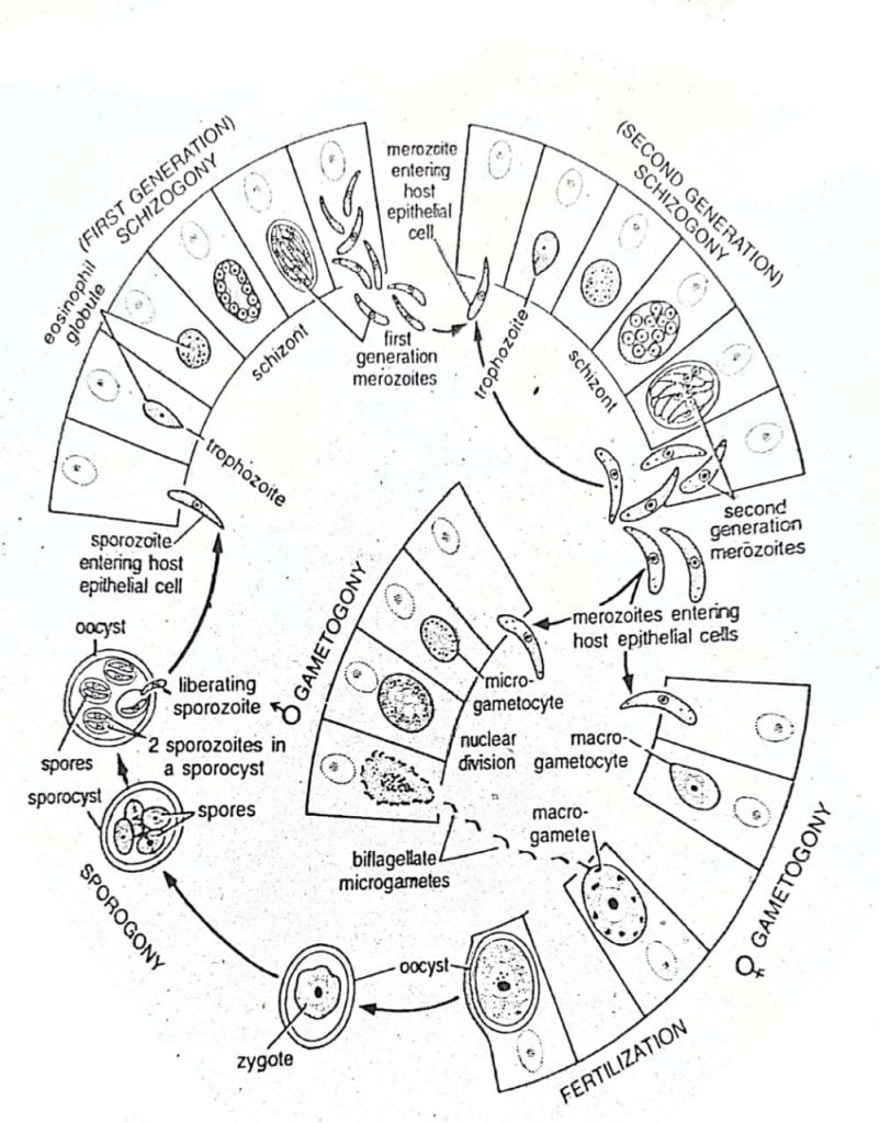
Fig: Life cycle of Eimeria tenella
I) Asexual Cycle (Schizogony)
In the life cycle of Eimeria tenella, there are two generations present in the schizogony: 1. First generation schizogony and 2. Second generation schizogony.
1. First Generation Schizogony
- This is the beginning of the life cycle of the Eimeria.
- It is initiated by the infection of epithelial cells of the caecum of the host by sporozoites.
The stages of the first generation schizogony (of the life cycle of Eimeria tenella) are described below.
a) Infection by sporozoites
-
-
- Oocysts or zygocysts are expelled out with the faecal matter of the infected fowl.
- If a healthy fowl swallow the oocysts, the wall of the oocysts breaks down and releases the sporozoites.
- The fowl’s digestive juice helps the oocyst break down the cyst wall.
- After releasing the sporozoites, the sporozoites enter the gut epithelial cell.
- The sporozoites then grow in size and multiply.
-
b) Sporozoite
-
-
- It is the earliest intracellular stage of the parasite.
- It is an elongated, slightly curved, microscopic, and unicellular organism.
- Its one end is pointed and the other end is blunt.
- Its external covering is called the pellicle.
- The pellicle contains contractile microtubules which help them in wriggling movement.
- The cytoplasm of the sporozoite contains a vesicular nucleus, a mitochondrion, endoplasmic reticulum, Golgi bodies, ribosomes, lysosomes, and reserve food.
- The roptries of the sporozoite secret the lytic secretion which help the organism penetrate.
-
c) Trophozoite
-
-
- The sporozoite grows continuously and becomes pear-shaped.
- This pear-shaped stage is called the trophozoite.
- An eosinophil globule is present at the blunt end of the trophozoite.
- It absorbs nutrients from the cytoplasm of the host cell through its external covering.
-
d) Schizont
-
-
- After the multiple fission of the trophozoite, it becomes the schizont.
- Repeated mitotic nuclear divisions have occurred which resulted in a multinucleate first-generation schizont.
- Somewhat oval in shape.
- The eosinophil globule persists in its cytoplasm.
-
e) Merozoites
-
-
- The schizont later turned into the merozoites.
- Almost 900 merozoites can be produced from a single first-generation schizont.
- They are very small, and inconspicuous and their length is 2-4 μ.
- Its one end is pointed and the other end is rounded.
- The pointed end contains a terminal granule and the rounded end contains a number of cytoplasmic granules.
- The first-generation merozoites later invade other epithelial cells.
-
2. Second Generation Schizogony
The liberated merozoites from the first generation schizogony then invade other epithelial cells and with this invasion, the second generation schizogony starts. The stages of the second generation schizogony (life cycle of Eimeria tenella) are given below-
a) Invasion of Fresh Host Cells
-
-
- The first-generation merozoite invades a fresh epithelial cell.
- The merozoite soon grows into a trophozoite.
-
b) Trophozoite
-
-
- The first generation trophozoite resembles the second generation trophozoite.
- It lacks an eosinophil globule.
- Its nucleus undergoes repeated mitotic divisions to form a multinucleate schizont.
-
c) Schizont
-
-
- There are so many differences between the first generation schizont and the second generation schizont.
- It is larger than the first generation schizont.
- The nuclei are scattered throughout its cytoplasm.
- Each nucleus contains a nucleolus.
-
d) Merozoite
-
-
- It is larger than the first-generation merozoites.
- It is elongated and pyriform.
- It has a rounded end and a blunt end.
- Within the schizont, each nucleus and some cytoplasmic matter around the nucleus form the second-generation merozoite.
- Each second-generation schizont forms about 250 second-generation merozoites.
- After releasing the merozoites from the schizont, they soon penetrate other epithelial cells of the caecum.
-
II) Sexual Cycle (Gametogony & Sporogony)
In the life cycle of Eimeria tenella, most of the second-generation merozoites do not repeat the asexual cycle. They start the sexual cycle. They develop into gametocytes which produce gametes.
1. Gametogony
- The gametocytes are of two types. Male or microgametocytes and female or macrogametocytes.
- Gametogony is an anisogamy type.
a) Microgametocytes
-
-
- After invading the host epithelial cells, some of the second-generation merozoites become microgametocytes immediately.
- They are small, oval and 5.5 – 18 μ in length.
- Later, it shows repeated nuclear divisions and forms microgametes.
- Microgametes are small, comma-shaped, and biflagellate.
- Each microgamete has a nucleus, mitochondrion, and two flagella.
-
b) Macrogametocytes
-
-
- After invading the host epithelial cells, some of the second-generation merozoites become macrogametocytes immediately.
- They are oval in shape.
- They are larger than the microgametocytes and measure about 8 – 25 μ in length.
- Each macrogametocytes later develop into a single macrogamete.
-
2. Fertilization
- Microgametes escape from the host cells and invade the cells which contain macrogametes.
- Microgametes and macrogametes fuse together and fertilization occurs.
- After the fertilization, it develops into a zygote which is diploid.
3. Oocyst
- The zygote secretes a thick double-layered cyst wall around itself when it is in the host cell.
- The outer layer is thick and the inner layer is delicate.
- It is oval in shape.
- It has no further development in the host cell.
- The oocysts come to the intestine by rupturing the host cell.
- From the intestine, the oocysts are expelled out to the environment.
4. Sporogony
- Within the oocysts, the zygotes divide by meiosis into four haploid cells, called the sporoblasts.
- Each sporoblast secretes a tough wall named sporocyst around itself and becomes a spore.
- Each spore further divides mitotically to produce two sporozoites.
5. Transference of Oocysts
- The infective oocysts are easily passed to another healthy fowl through direct ingestion of faecal matters.
6. Liberation of Sporozoites
- After the ingestion of faecal matters by a healthy fowl, the cyst wall breaks down with the help of digestive enzymes like pepsin and trypsin and the sporozoites got released. Thus the life cycle of Eimeria tenella completes.
———THE END———
See more:
- Life Cycle of Leishmania donovani | Diagram
- Life Cycle of Trypanosoma gambiense | Diagram
- Life Cycle of Ascaris lumbricoides | Diagram
- General Characters of All Phylum of The Invertebrates.
Reference:
1. Eimeria wiki

Md Ekarm Hossain Bhuiyan is a dedicated zoology graduate with a profound passion for the study of animal life. He completed his primary and secondary education at Ispahani Public School and College, renowned for its commitment to academic excellence. He then pursued his secondary education at Government Science College. After that he achieved graduation at Department of Zoology, Jagannath University. His educational background and enthusiasm for zoology position him to make meaningful contributions to the field of biological sciences in Bangladesh.