In this article, we will learn the life cycle of Ascaris lumbricoides.
What is the life cycle?
The life cycle refers to the sequence of developmental stages that an organism goes through from its initial formation to its reproduction. It includes the process of growth, development, and reproduction. The different stages in the life cycle of an organism can vary depending on the species, but they usually include a period of growth and development, followed by reproduction and the creation of new individuals. These stages can be further divided into sub-stages, such as the embryonic stage in animals, or the germination stage in plants. The life cycle is a fundamental aspect of the biology of an organism and can be used to understand its behavior, ecology, and evolution.
Life Cycle of Ascaris lumbricoides
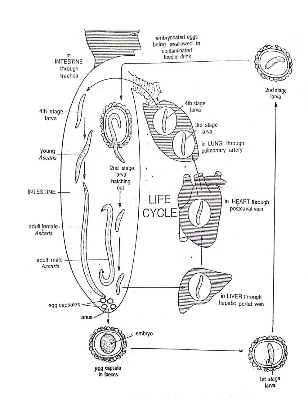
Fig: Life Cycle of Ascaris lumbricoides
- In the life cycle of Ascaris, there is no secondary host. So, the life cycle is monogenetic as it involves only one host.
- Man is the only host of Ascaris.
- The life cycle includes several steps such as copulation and fertilization, zygote, cleavage and early development, infection of a new host, and later development and migration.
The steps are described below.
1. Copulation and Fertilization
- Copulation takes place in adult worms.
- It takes place in the small intestine of a man.
- The male orients its body at right angles to that of the female in such a way that its cloacal aperture apposes the vulva of the female.
- The penial setae of male help to open the vulva and sperms are transferred into the vagina.
- Then the sperms pass on to the proximal ends of the uteri.
- Fertilization takes place in the proximal ends of uteri or distal ends of oviducts.
- It is caused by the entry of a sperm into the ova.
- After fertilization, the ova undergo second maturation division.
2. Zygote
- Unfertilized eggs contain globules of glycogen and fat.
- Glycogen globules migrate to the surface and form a fertilization membrane immediately after fertilization.
- The fertilization membrane hardens into a thick, clear, and chitinous shell.
- Then the fat globules form a thin layer below the shell.
- As the fertilized egg or zygote moves down the uterus, the uterine wall secretes around it a thick and hard coat.
- Fertilized egg or zygote is usually measuring around 50-70 micrometers in diameter, and it is typically spherical in shape.
- The uteri of a single mature female may contain as many as 27 million eggs.
- The zygote of Ascaris lumbricoides is formed within the female worm and is released into the environment when the female lays eggs.
- The eggs are then passed out of the body of the infected individual through faeces.
- The eggs can survive in the environment for weeks to months, and they become infective when they come in contact with suitable conditions such as moisture and warmth.
- Once the eggs are ingested by a human host, they hatch within the small intestine. They mature into adult worms within the small intestine, where they can live for up to 2 years.
3. Cleavage and Early Development
- Cleavage is the process of cell division that occurs during the early stages of development in animals.
- The cleavage is a type of spiral cleavage.
- The first division results in a dorsal and a ventral cell.
- The dorsal cell divides into an anterior and a posterior cell. The ventral cell divides into an upper and a lower cell. Thus the four-celled embryo is formed.
- The initial cleavage divisions are rapid and synchronous, resulting in a ball of cells called a morula.
- The morula then develops into a blastula, which is a 16-celled embryo.
- As the cleavage continues, the blastula develops into a gastrula, which is the early stage of the worm’s body plan.
- The gastrula then develops into a juvenile in 10-14 days from the start of cleavage.
- This juvenile is also termed as rhabditoid larva of the first stage. It is not capable of infecting the host.
- After a week, it moults within the eggshell and becomes the second stage of rhabditoid larva, which is capable of infecting the host.
- Under suitable conditions, the infective eggs remain viable for about six years.
4. Infection of New Host
- Man acquires infection by directly ingesting eggs containing the infective second-stage rhabditoid larva.
- The eggs are typically acquired through the ingestion of contaminated food or water.
- In the small intestine, man’s digestive juice dissolves the egg shells and the juveniles hatch out.
- Symptoms of the infection can include abdominal pain, diarrhea, and malnutrition, and in some cases, the worms can even migrate to the lungs, causing cough and difficulty breathing.
5. Later Development and Migration
- After hatching, the juvenile performs active thrashing movements and bores through the epithelium of the host’s intestine and starts its migration into the host’s body.
a) Primary Migration
-
-
- The larva enters into the hepatic portal circulation.
- The hepatic portal circulation carries the larva to the liver.
- From the liver, the larva reaches the heart through post-caval vein.
- From the heart, it is transported to the lung via the pulmonary artery.
- It stays in the lungs for a few days.
- Here the larva moults 2 times to grow into the fourth stage larva.
-
b) Secondary Migration
-
-
- In the fourth stage, the larva leaves the lungs and reaches the pharynx.
- Then it is again swallowed into the gut.
- In the intestine, it becomes an adult.
-
c) Aberrant Migration
-
-
- Sometimes larva of Ascaris does not follow the usual path but reaches the brain or spinal cord or any such organ.
- The larva is not able to survive in these organs.
-
———— THE END ————-
Read More:
- Life Cycle of Eimeria tenella – With Diagram
- General Characters of All Phylum of The Invertebrates.
- Ascariasis – CDC

Md Ekarm Hossain Bhuiyan is a dedicated zoology graduate with a profound passion for the study of animal life. He completed his primary and secondary education at Ispahani Public School and College, renowned for its commitment to academic excellence. He then pursued his secondary education at Government Science College. After that he achieved graduation at Department of Zoology, Jagannath University. His educational background and enthusiasm for zoology position him to make meaningful contributions to the field of biological sciences in Bangladesh.