The digestive system of Columba livia, also known as the rock pigeon, is similar to that of other birds. Food enters the mouth and is mashed by the beak and tongue before being swallowed and entering the oesophagus, stomach, small intestine and large intestine. Nutrients are absorbed into the bloodstream through the walls of the small intestine, and indigestible material is passed into the large intestine and then eliminated through the cloaca.
What is Digestive System?
The digestive system is a series of organs that work together to convert food into energy and basic nutrients to feed the entire body. Food is broken down through a mechanical and chemical process and then absorbed into the bloodstream to be transported to cells throughout the body.
Digestive System of Columba livia
- The digestive system of pigeon includes the alimentary canal and the digestive glands.
- Due to the loss of teeth and arial mode of life, the digestive system of pigeon like other birds is highly modified.
- The alimentary canal is short in comparison to the size of the bird.
- Moreover, its different parts are modified in such a way, that assimilation of digested food takes no time.
The whole digestive system of pigeon is divided into two parts:
1. The alimentary canal and
2. The digestive glands.
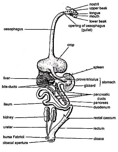
Alimentary Canal
- It is a long coiled tube.
- It begins at the mouth.
- It comprises of mouth, buccal cavity, pharynx, oesophagus, stomach, small intestine, large intestine, and cloacal aperture.
- It ends at the cloaca.
A. Mouth
-
-
- The alimentary canal starts here.
- It is a wide slit bounded by upper and lower horny beaks.
- Teeth are absent.
- The mouth opens into the buccal cavity.
-
B. Buccal Cavity
-
-
- The interior of the buccal cavity is comparatively featureless.
- The tongue is present on the floor of the buccal cavity.
- The tongue is large, narrow, somewhat triangular, and pointed at the tip.
- The anterior pointed end of the tongue is free.
- Some taste buds and numerous mucous glands are present on the surface of the tongue.
-
C. Pharynx
-
-
- The posterior part of the buccal cavity may be termed the pharynx.
- A pair of apertures, the posterior nares, open on its roof. They are covered by palatal folds of the skull roof.
- Behind the nares opens the single median aperture of the eustachian or pharyngotympanic tubes.
- Each tube extends to the cavity of the middle ear.
- Near the base of the tongue, at the floor of the pharynx, there is an aperture called glottis is present which leads into the trachea.
-
D. Oesophagus and Crop
-
-
- The pharynx leads into the oesophagus.
- It is a long, wide, distensible and thick-walled tube.
- It runs back through the neck to join the stomach.
- It expands into a thin-walled, bilobed, elastic sac called the crop at the base of the neck.
- The crop serves as a food reservoir.
- The dry and hard food grains are stored, moistened and softened at the crop.
- The pigeon has a unique ability to produce “pigeon’s milk” which is nourishing secretion from both sexes during the breeding season. This milk is formed by the degeneration of the epithelial cells lining the crop.
- Behind the crop, the oesophagus again becomes thick-walled and enters the stomach.
-
E. Stomach
-
-
- The stomach of the pigeon the divided into two parts: proventriculus and gizzard.
-
Proventriculus
-
-
-
-
- The proventriculus is a small but thick-walled sac, externally appearing like a slight dilation of the oesophagus.
- The gastric juice is secreted here.
- The spleen is attached to the right side of the proventriculus with the peritoneum.
-
-
-
Gizzard
-
-
-
-
- The gizzard is the large, hard and laterally compressed part of the stomach.
- It is biconvex lens shaped.
- The gizzard represents the pyloric region of the stomach.
- The cavity of gizzard always contains small stones called gastroliths swallowed by the bird. These stones help the gizzard in grinding up the food.
- The opening of the gizzard into the small intestine is guarded by the pyloric valve or pylorus or pyloric sphincter.
-
-
-
F. Small Intestine
-
-
- It is about 75 cm long and is divided into two parts: duodenum and ileum.
-
Duodenum
-
-
-
-
- The gizzard opens into the duodenum.
- It is the anterior part of the small intestine.
- The duodenum forms a U-shaped loop enclosing the pancreas between its two limbs.
- Internally duodenum contains villi, crypts of Liberkuhn and goblet cells.
- The ducts from the pancreas and lobes of the liver open into the duodenum thus the pancreatic juice and bile are released into the duodenum.
-
-
-
Ileum
-
-
-
-
- It is the second part of the small intestine.
- It is a very long and extensively convoluted tube.
- Its inner lining has numerous finger-like villi.
- These villi greatly increase the area of secretion and absorption of the ileum.
-
-
-
G. Large Intestine
-
-
- The ileum opens into the large intestine.
- The junction of the ileum and large intestine bears a pair of small pouches named rectal or colic caeca.
- The large intestine is differentiated into two parts: rectum and cloaca.
-
Rectum
-
-
-
-
- It is a narrow anterior part of the large intestine.
- It is about 4 cm long.
- Its opening into the cloaca is guarded by an anal sphincter.
-
-
-
Cloaca
-
-
-
-
- It is the terminal part of the alimentary canal.
- Rectum opens into a chamber called the cloaca.
- The cloaca is differentiated into three chambers: coprodaeum, urodaeum and proctodaeum.
- Coprodaeum receives the rectum, urodaeum receives the ureters and genital ducts and the proctodaeum opens to the outside through the cloacal aperture or vent.
- It is the common releasing pathway for faeces, urine and genital products.
-
-
-
Digestive Glands
Approximately 8 digestive glands are found in a pigeon’s body and they are:
A. Buccal Glands
-
-
- These glands are present in the buccal cavity.
- They mainly secrete mucus to moisten food and probably amylase.
- They are not salivary.
-
B. Salivary Glands
-
-
- Paired angular and unpaired sublingual salivary glands are found in the pharyngeal region.
- These glands secrete saliva which moistens the food.
-
C. Gastric Glands
-
-
- The thick epithelial lining of the proventriculus contains numerous gastric glands.
- Gastric glands are large and they are visible to the naked eye.
- These glands secrete gastric juice.
-
D. Liver
-
-
- It is a large, dark red and bilobed gland.
- The lobes are called the right lobe and left lobe. The right lobe is larger than the left.
- There are two bile ducts, one from each liver lobe.
- The left bile duct opens into the proximal limb and the right bile duct opens into the distal limb of the duodenum.
- There is no gall bladder in the pigeon’s liver.
- The liver secretes bile.
-
E. Pancreas
-
-
- The pancreas is a compact reddish gland.
- It is located between the two limbs of the duodenum.
- There are three pancreatic ducts, all opening into the distal limb of the duodenum.
- It secretes pancreatic juice which contains several enzymes.
-
F. Intestinal Glands
-
-
- These glands are located inside the small intestine.
- They secrete various enzymes.
-
G. Tubular Glands
-
-
- The internal lining of the gizzard contains these glands.
- The fluid secreted by these glands is thick, horny and yellowish-green in colour.
-
H. Caecal Glands
-
-
- These glands produce digestive juices.
- These glands are also helpful in the absorption of water from food.
-
——–THE END——–
Read More:
- Air Sacs of Pigeon | Bird | Columba livia | Diagram | Note
- Muscular System of Columba livia | Pigeon | Diagram
- Feathers in Pigeon | Columba livia | Diagram
- Skin and Exoskeleton of Columba livia | Pigeon | Diagram
- External Morphology of Columba livia | Pigeon | Diagram
- General Characters of All Classes of Vertebrates.
- The Sound Producing Organ in Birds | Syrinx | Diagram | Note
Reference:

Md Ekarm Hossain Bhuiyan is a dedicated zoology graduate with a profound passion for the study of animal life. He completed his primary and secondary education at Ispahani Public School and College, renowned for its commitment to academic excellence. He then pursued his secondary education at Government Science College. After that he achieved graduation at Department of Zoology, Jagannath University. His educational background and enthusiasm for zoology position him to make meaningful contributions to the field of biological sciences in Bangladesh.