Cavia porcellus is the scientific name of Guinea pig. In this article, we will learn about the circulatory system of Cavia porcellus or Guinea pig. They are mammals under the Rodentia order.
Circulatory System of Cavia
Two circulatory fluids are present and they are blood and lymph. Blood is circulated to the whole body through the well-defined vessels and it is pumped by the heart. The lymph flows through the lymph vessels.
Blood Circulatory System of Cavia
The blood circulatory system of Cavia consists of blood, heart, and blood vessels.
1. Blood
-
-
- The blood of Cavia consists of liquid plasma and some corpuscles.
- The color of the blood plasma is pale yellow.
- The liquid plasma contains various inorganic salts, vitamins and hormones.
- Three different types of blood corpuscles are present in the blood of Cavia. They are erythrocytes, leucocytes and thrombocytes.
- Mature erythrocytes or red blood corpuscles or RBCs are round, non-nucleated and contain a red pigment named haemoglobin. As the color of haemoglobin is red, the color of the blood of Cavia is red.
- The erythrocytes carry oxygen to the whole body and carry back carbon dioxide from the whole body to the lungs.
- Leucocytes or White Blood Corpuscles or WBCs are much larger than RBCs and much less in number.
- Leucocytes move in an amoeboid fashion and they act as the scavenger of the body.
- Thrombocytes or Platelets are also non-nucleated like RBCs.
- Thrombocytes prevent the loss of blood from the body if the body is injured.
-
2. Heart
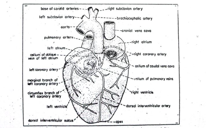
fig : Heart of Guinea pig.
-
-
- The heart of the Cavia is a muscular, hollow, four-chambered, and cone-shaped organ.
- The heart is located in a space within the thorax between the two pleural bags.
- The name of the space is mediastinum where the heart is located.
- The covering of the heart is the pericardium. It is a thin peritoneal membrane.
- The pericardium is two-layered. The outer layer of the pericardium is fibrous pericardium and the inner layer of the pericardium is the serous pericardium.
- The serous pericardium is again divided into two layers. The outer layer is called the parietal layer and the inner layer is called the visceral layer.
- There is a space between the fibrous layer and the serous layer. The space is called the pericardial cavity.
- The heart has two auricles and two ventricles.
-
A. Right Auricle
-
-
-
-
- The right auricle lies cranial to the right ventricle.
- The right auricle receives the venous systemic blood.
- An internally located ridge divides the right auricle into two regions. The regions are 1. Sinus venarum cavarnum and 2. Right auricula.
- There are four openings present in the right auricle.
- The blood enters the right auricle through three openings. They are the Coronary sinus, the Larger ostium of caudal vena cava and the Smaller ostium of the cranial vena cava.
- With the other opening, the blood passes on to the right ventricle from the right auricle and the name of the opening is right atrioventricular ostium.
-
-
-
B. Right Ventricle
-
-
-
-
- The right ventricle lies caudal to the right auricle.
- It has thick walls.
- It receives blood from the right auricle through the right atrioventricular ostium.
- The ostium is surrounded by the right atrioventricular valve and the valve is known as the tricuspid valve because the valve is formed by three cusps.
- The pulmonary aorta arises from the right ventricle. The right ventricle supplies blood to the lungs through the pulmonary aorta.
-
-
-
C. Left Auricle
-
-
-
-
- It is smaller in size than the right auricle.
- It is separated internally from the right auricle by the inter-auricular septum.
- It has left auricula internally which is similar to the right auricula.
- It receives blood through the pulmonary vein.
- The pulmonary vein is present on the dorsal side of the left auricle.
- The left auricle opens to the left ventricle through the left atrioventricular ostium.
-
-
-
D. Left Ventricle
-
-
-
-
- It is larger than the right ventricle.
- It has thick walls.
- The left atrioventricular ostium is provided with a bicuspid valve which has two unequal cusps.
- The larger cusp of the bicuspid valve is the septal cusp and the smaller one is known as the parietal cusp.
- The left aortic arch arises from it.
- The left ventricle opens to the left aortic arch through the aortic ostium.
- The aortic ostium is guarded by the aortic valves which have three semilunar cusps.
- The valves prevent the backflow of the blood.
-
-
-
3. Blood Vessels
-
-
- The blood is supplied from the heart to the different parts of the body through the blood vessels.
- Cavia has well-developed blood vessels.
- The circulatory system of Cavia is a closed-type circulatory system because the blood of the Cavia remains enclosed in the blood vessels.
- Oxygenated blood is carried away from the heart to the different parts of the body through the arteries (except the pulmonary artery).
- Deoxygenated blood is collected from the different parts of the body by the veins (except the pulmonary vein) and carried to the lungs.
- The arteries break up into arterioles.
- The arterioles again break up into arterial capillaries.
- The arterial capillaries unite with the venous capillaries and form a capillary network.
- Venous capillaries unite to form venules.
- Venules unite to form veins.
- Thus a closed type circulatory system is formed.
-
Mechanism of Blood Circulation through Heart
- The heart works day and night continuously.
- The contraction of the heart is called systole and the relaxation of the heart is called diastole.
- Both auricles begin their systole and diastole at the same time. The ventricles also perform their systole and diastole at the same time.
- The two auricles filled with blood because of the diastole.
- The right auricle receives deoxygenated blood from the venous system and the left auricle receives oxygenated blood from the pulmonary vein.
- Then the two auricles begin their systole at the same time.
- Blood from the auricles is forced to the ventricles.
- Valves located between the auricles and ventricles prevent the backflow of the blood.
- After filling up with the blood, the ventricles start systole.
- Then, blood from the right ventricle is forced to the pulmonary artery and blood from the left ventricle is forced to the aortic arch.
- Here, the valves located between the arteries and the ventricles prevent the backflow of the blood.
- When the auricles start systole, the ventricles start diastole and when the ventricles start systole, the auricles start diastole.
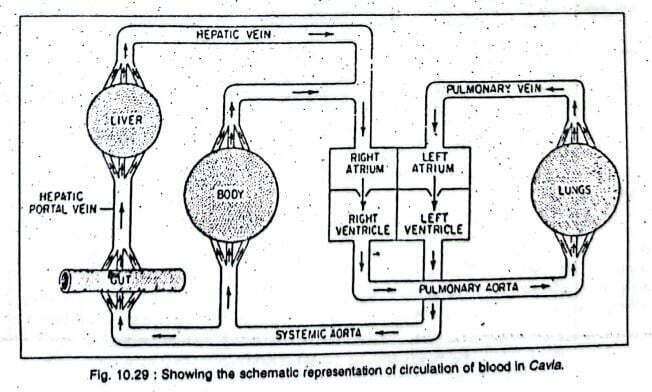
Scheme of Blood Circulation
————-THE END—————
Read More:
- Respiratory System of Guinea Pig | Cavia porcellus | Diagram
- Nervous System of Guinea Pig | Cavia porcellus | Diagram
- Digestive System of Guinea Pig | Cavia porcellus | Diagram
- External Morphology of Guinea Pig | Cavia porcellus | Diagram
- General Characters of All Classes of Vertebrates.
Reference:

Md Ekarm Hossain Bhuiyan is a dedicated zoology graduate with a profound passion for the study of animal life. He completed his primary and secondary education at Ispahani Public School and College, renowned for its commitment to academic excellence. He then pursued his secondary education at Government Science College. After that he achieved graduation at Department of Zoology, Jagannath University. His educational background and enthusiasm for zoology position him to make meaningful contributions to the field of biological sciences in Bangladesh.