Hirudinaria is a genus of medicinal leeches that belong to the phylum Annelida. These leeches are hermaphroditic and possess a complex digestive system adapted for their blood-feeding lifestyle. In this article, we will learn about the digestive system of Hirudinaria.
Diagram of The Digestive System
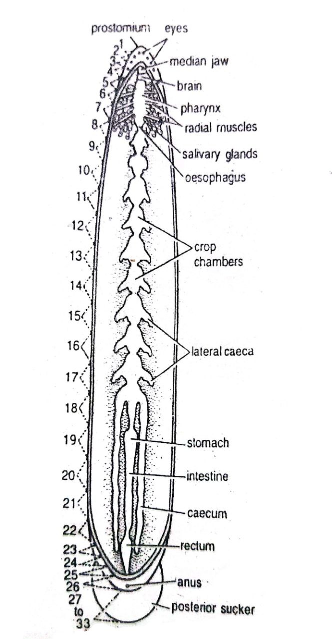
The Digestive System of Hirudinaria
The digestive system of Hirudinaria or leech is described below in two parts: the alimentary canal and food, feeding, and digestion mechanism.
Alimentary Canal of Leech
- The alimentary canal of the leech is complete.
- It is a straight tube.
- The alimentary canal starts from the mouth and ends at the anus.
- It is divided into the buccal cavity, pharynx, oesophagus, crop, stomach, intestine, and rectum.
1. Pre-Oral Chamber and Mouth
-
-
- The pre-oral chamber is a cup-like depression.
- The pre-oral chamber is located at the ventral side of the oral sucker or anterior sucker.
- The mouth is located on the roof of the pre-oral chamber.
- The roof of the pre-oral chamber is formed by a membrane-like structure called the velum.
- The mouth is a triradiate opening, located at the center of the velum.
-
2. Buccal Cavity
-
-
- The mouth leads into the buccal cavity.
- It is a short chamber behind the velum.
- Three crescentic jaws are present.
- One jaw is mid-dorsal and the other two jaws are ventro-lateral in position.
- Each jaw bears minute teeth or denticles in a single row.
- The number of teeth is 103-128 on the median jaw and 85-115 on each lateral jaw.
- Each jaw contains numerous openings of salivary glands.
- Jaws produce the triradiate bite or Y-shaped wound in the skin of the host.
-
3. Pharynx
-
-
- The buccal cavity leads into the pharynx.
- The pharynx of leech is highly muscular.
- It is an oval sac extending from the 5th-8th segments.
- Its inner surface bears longitudinal folds.
- Externally, it is surrounded by salivary glands.
- Secretion of these glands contains hirudin or anticoagulin.
- The hirudin or anticoagulin prevents the coagulation of blood while the leech is feeding.
-
4. Oesophagus
-
-
- The pharynx leads into the oesophagus.
- It is a short and narrow tube.
- It has a very narrow lumen and a much folded epithelial lining.
-
5. Crop
-
-
- The oesophagus leads into the crop.
- It is the largest portion of the alimentary canal.
- It extends from the 9th to the 18th segment.
- The crop is capable of great dilation to store an enormous quantity of blood.
-
6. Stomach
-
-
- The last chamber of the crop leads into a funnel-shaped tube.
- The tube leads into a small heart-shaped stomach.
- The stomach is present in the 19th segment.
- The mucous lining of the stomach is thrown into anastomosing transverse folds.
-
7. Intestine
-
-
- The stomach leads into the intestine.
- It is a thin-walled, straight, and narrow tube.
- It extends from the 20th to the 22nd segments.
- Its inner lining is thrown into numerous spiral folds and villi-like processes which increase its absorptive surface.
-
8. Rectum
-
-
- The intestine is followed by the rectum.
- It is a somewhat dilated and thin-walled tube.
- It extends from the 23rd to the 26th segments.
- It opens to the exterior through the anus.
- The anus is present mid-dorsally on the 26th segment.
-
————-THE END————–
Read More:
- External Morphology of Hirudinaria granulosa | Leech | Diagram
- General Characters of All Phylum of The Invertebrates.

Md Ekarm Hossain Bhuiyan is a dedicated zoology graduate with a profound passion for the study of animal life. He completed his primary and secondary education at Ispahani Public School and College, renowned for its commitment to academic excellence. He then pursued his secondary education at Government Science College. After that he achieved graduation at Department of Zoology, Jagannath University. His educational background and enthusiasm for zoology position him to make meaningful contributions to the field of biological sciences in Bangladesh.