
Structure of Human Heart
Human heart is divided into four chambers. Two atria and two ventricles. The upper two chambers are the right and left atria and the lower two chambers are the right and left ventricles. So basically the four chambers are
- Right atrium
- Right ventricle
- Left atrium
- Left ventricle
The ventricular walls are more thicker than the walls of the atria. The left ventricular wall is three times thicker than the right ventricular wall. The inter atrial-septum divides the two atria and the inter ventricular-septum divides the two ventricles.
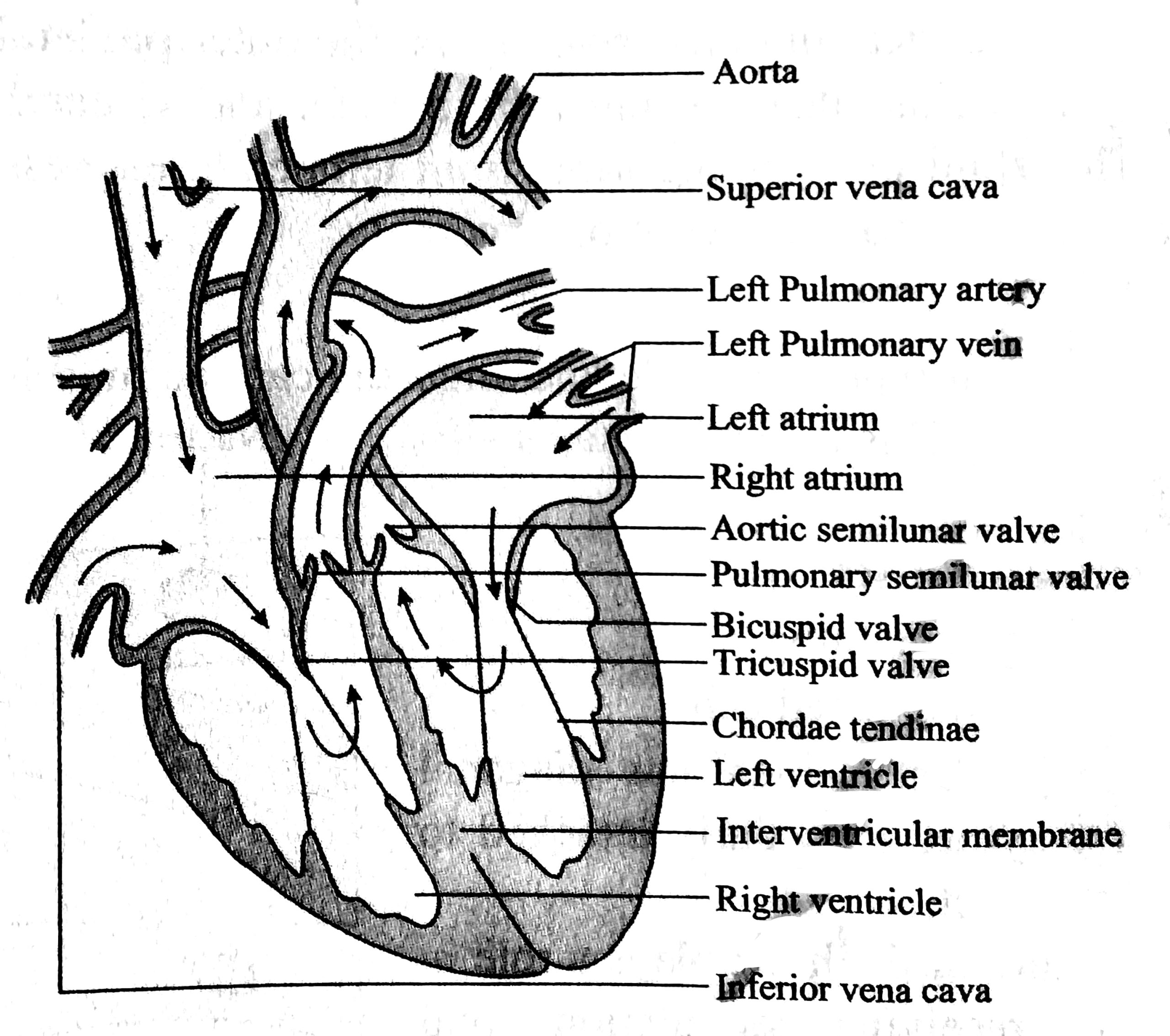
The details about the four chambers of the heart is given below:-
1. Right Atrium
- It is comparatively larger than the left atrium.
- It is made of thin wall.
- Sino-atrial node (a pacemaker) is situated in the right atrium.
- Heartbeat is originated from sino-atrial node (SAN).
- Blood from anterior side of the body comes to the right atrium through superior venacava.
- Blood from posterior side of the body comes to the right atrium through inferior venacava.
- The right atrium collects carbon di oxide rich blood only.
- Tricuspid valves are present between the right atrium and right ventricle.
2. Right Ventricle
- The right ventricle is a little larger than the left ventricle.
- It collects the carbon di oxide rich blood from the right atrium.
- The blood travels to the lungs from the right ventricle through pulmonary artery.
- Pulmonary artery is the only artery which carries carbon di oxide rich blood.
- Semilunar valves are present between the right ventricle and pulmonary artery. The valves prevent the backward flow of the blood.
- The semilunar valves are also known as pulmonary valves.
3. Left Atrium
- It is smaller than the right atrium.
- The walls are comparatively thicker than the right atrium.
- It collects oxygenated blood from the lungs through pulmonary vein.
- Pulmonary vein is the only vein in the body which carries oxygenated blood.
- An opening is present between the left atrium and left ventricle.
- This opening is guarded by bicuspid valves.
4. Left Ventricle
- The wall of the left ventricle is the thickest.
- It collects oxygenated blood from the left atrium.
- Left ventricle pumps the oxygenated blood to the whole body through aorta.
- There is a semilunar valve between the left ventricle and the aorta. This valve is also known as aortic valve.
Read More:
- Structure of Euglena with Labeled Diagram
- Structure & Diagram of Amoeba proteus
- Structure, Development &; Homology of Placoid Scales | Shark | Scoliodon

Md Ekarm Hossain Bhuiyan is a dedicated zoology graduate with a profound passion for the study of animal life. He completed his primary and secondary education at Ispahani Public School and College, renowned for its commitment to academic excellence. He then pursued his secondary education at Government Science College. After that he achieved BSc. in Zoology and MSc. in Entomology at Department of Zoology, Jagannath University, Dhaka. Currently he is teaching zoology at Milestone College, Dhaka. His educational background and enthusiasm for zoology position him to make meaningful contributions to the field of biological sciences in Bangladesh.