Amoeba proteus is a single-celled organism. It is a eukaryotic organism, which means that it has a true nucleus and other membrane-bound organelles. Amoeba proteus is a freshwater organism that is commonly found in ponds, lakes, and streams. It is also widely used as a model organism in biological research. This organism has a distinctive shape, with a flexible and amorphous body that constantly changes shape as it moves. It uses pseudopodia, or false feet, to move and capture food.
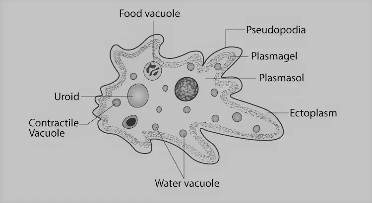
Diagram of Amoeba proteus
Structure of Amoeba proteus
1. Shape and Size
- It is a unicellular, microscopic animalcule.
- It measures about 250-600 µ.
- To the naked eye, the larger Amoeba proteus is visible as a whitish blob.
- It appears as an irregular, colorless, and translucent mass of protoplasm under the microscope.
- It can continuously change its shape.
- It has a definite anterior and posterior end.
- At the anterior end, the animal puts out pseudopodia.
- The posterior end is marked by a wrinkled region called uroid.
2. Pseudopodia
- Pseudopodia are also called false feet of Amoeba.
- They are the irregular blunt processes of the cell body.
- These pseudopodia are of variable size.
- These are capable of protruding or retracting.
- As many pseudopodia are formed simultaneously, Amoeba proteus are called ‘Polypodial’ species.
- Its pseudopodia are large and broad with rounded tips. Such pseudopodia are called lobopodia.
- These pseudopodia assist the animal in locomotion and food ingestion.
3. Plasmalemma
- Amoeba possesses no pellicle or cell wall.
- The body is covered by a thin, delicate plasma membrane which is called plasmalemma.
- This membrane is selectively permeable.
- Water and some small solute molecules can pass freely through it.
- The plasmalemma has numerous fine, ridge-like extensions on its outer surface.
4. Cytoplasm
- Inside the plasmalemma, there is a dense mass of cytoplasm containing various organelles.
- The cytoplasm is divided into two zones and they are ectoplasm and endoplasm.
a) Ectoplasm
-
-
- It is the outer zone of the cytoplasm.
- It lies immediately beneath the plasmalemma.
- It is thin, clear and transparent.
- It is somewhat rigid, contractile and under tension.
- It is most clearly visible at the tip of a pseudopodium where it forms a hyaline cap.
-
b) Endoplasm
-
-
- It is the inner zone of the cytoplasm.
- It is completely surrounded by ectoplasm.
- It is fluid-like, granular and semi-transparent.
- As the ectoplasm is under tension, the endoplasm must also be under the pressure of the ectoplasm.
-
5. Endoplasmic Organelles
a) Nucleus
-
-
- There is a single, large, flattened and slightly biconcave nucleus.
- It can be present anywhere in the endoplasm.
- It is bounded by a thin nuclear membrane.
- A honeycomb-like lattice is found below the inner nuclear membrane.
- The nucleoplasm is a clear substance with scattered chromatin granules.
-
b) Contractile Vacuole
-
-
- A single, clear and rounded contractile vacuole is present at the posterior end of the endoplasm.
- It is filled with watery fluid and enclosed by a unit membrane.
- It helps in the osmoregulatory and excretory activities of the animal.
-
c) Food Vacuoles
-
-
- A number of spherical food vacuoles are present in the endoplasm.
- They contain water and food in various phases of digestion.
- These are formed when Amoeba engulfs food with a drop of water.
- Digestion takes place inside these food vacuoles.
- They disappear with the egestion of non-digestible food from the body.
-
d) Water Globules
-
-
- These are several, small and spherical vacuoles filled with a colorless fluid.
-
e) Other Organelles
-
-
- The endoplasm contains endoplasmic reticulum which forms a network of tubules.
- The endoplasm also contains ribosomes that occur on some of the endoplasmic vesicles.
- The Golgi bodies are present as several groups of sac-like tubes.
- The mitochondria are more or less oval and have tubular cristae.
- The lysosomes are membrane-bound spherical bodies, scattered in the endoplasm.
-
———– THE END ———–
Read More:
- Locomotion or Movement of Amoeba proteus | Diagram
- General Characters of All Phylum of The Invertebrates.

Md Ekarm Hossain Bhuiyan is a dedicated zoology graduate with a profound passion for the study of animal life. He completed his primary and secondary education at Ispahani Public School and College, renowned for its commitment to academic excellence. He then pursued his secondary education at Government Science College. After that he achieved graduation at Department of Zoology, Jagannath University. His educational background and enthusiasm for zoology position him to make meaningful contributions to the field of biological sciences in Bangladesh.