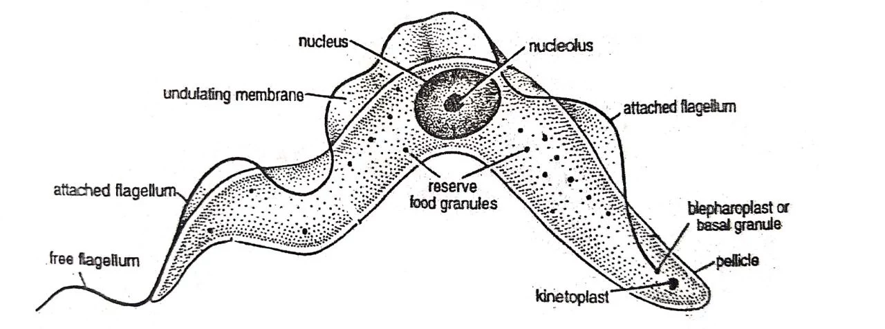
The basic morphology of Trypanosoma gambiense is that of a single-celled organism, known as a protozoan. It has a characteristic elongated, cigar-shaped body, with a distinct anterior and posterior end. The body is surrounded by a plasma membrane, which encloses the cytoplasm and organelles. In this article, we will learn the morphology of Trypanosoma gambiense.
Morphology of Trypanosoma gambiense

1. Shape and Size
-
-
- Trypanosoma gambiense is unicellular, microscopic, elongated, leaf-like, flattened, and tapering at both sides.
- Its length is about 10-40µ and its width is about 2.5-10µ.
- The anterior end is pointed and the posterior end is blunt.
- It is a polymorphic species and occurs in two developmental forms, the trypanosome form, and the crithidial form.
- In the bloodstream of man, the trypanosome form may be long slender or short stumpy, or intermediate between the two.
- In the tsetse fly, the trypanosome form is long and slender in the midgut, and short and stumpy in the salivary gland.
- The crithidial form is represented only in the salivary glands of the tsetse fly.
-
2. Pellicle
-
-
- The whole body is surrounded externally by pellicle.
- The pellicle is a thin and elastic covering.
- It is supported by the fine fibrils, called microtubules.
- Microtubules may help to maintain the shape of the organism while it swims in the blood plasma.
-
3. Flagellum
-
-
- Trypanosoma gambiense is uniflagellate as it has a single flagellum.
- The flagellum arises from the posterior part of the body.
- It runs forward, attached along the entire length of the body, and becomes free at the anterior end of the body.
- When the trypanosome moves in blood, the undulating waves pass from the tip to the base of the flagellum.
-
4. Undulating Membrane
-
-
- When the flagellum beats, the pellicle, to which it is attached, is pulled up into a membranous fold called the undulating membrane.
- It may be an adaptation for locomotion in fluid like blood.
-
5. Cytoplasm
-
-
- The cytoplasm is enclosed within the pellicle.
- The cytoplasm is not differentiated into ectoplasm and endoplasm.
- An elongated mitochondrion with tubular cristae is present in the cytoplasm.
- Just near the basal body of the flagellum (at the posterior end of the body), the mitochondrion forms a disk-like structure called the parabasal body or kinetoplast.
- The kinetoplast includes a double-stranded DNA helix.
- A Golgi apparatus, endoplasmic reticulum, and ribosome are present in the cytoplasm.
- The cytoplasm also contains numerous greenish granules called voluntin granules. These are supposed to be stored food particles.
- Various small vacuoles are also seen in the cytoplasm, some with hydrolytic enzymes.
-
6. Nucleus
-
-
- The nucleus is present within the cytoplasm.
- The nucleus is large, oval, and vesicular.
- The nuclear membrane is a double-unit membrane and bears pores.
- The nucleolus is present at the center of the nucleus and surrounded by a clear space.
- Numerous chromosomes are present within the nucleoplasm.
-
———— THE END ———–
Read More:
- African Sleeping Sickness or Trypanosomiasis: Symptoms, Diagnosis, Treatment, and Prevention
- Life Cycle of Trypanosoma gambiense | Diagram
- General Characters of All Phylum of The Invertebrates.
Reference:
- “Modern Textbook of Zoology Invertebrates” written by R. L. Kotpal.
- CDC – African Trypanosomiasis – Biology

Md Ekarm Hossain Bhuiyan is a dedicated zoology graduate with a profound passion for the study of animal life. He completed his primary and secondary education at Ispahani Public School and College, renowned for its commitment to academic excellence. He then pursued his secondary education at Government Science College. After that he achieved graduation at Department of Zoology, Jagannath University. His educational background and enthusiasm for zoology position him to make meaningful contributions to the field of biological sciences in Bangladesh.