Duttaphrynus melanostictus or Bufo melanostictus is the scientific name of Asian common toad. In this article on Duttaphrynus melanostictus or Bufo melanostictus, we will learn 1. Systematic position of Asian common toad, 2. Special features of Bufo melanostictus, 3. External morphology of Duttaphrynus melanostictus.
Systematic Position of Asian Common Toad
Kingdom: Animalia
Phylum: Chordata
Class: Amphibia
Order: Anura
Family: Bufonidae
Genus: Duttaphrynus
Species: Duttaphrynus melanostictus
Special Features of Asian Common Toad
1. They have a wide distribution across South and South East Asia.
2. They are bilaterally symmetrical.
3. Their skin is naked and rough and they have parotid glands for their defense.
4. Vertebral column is present.
5. The tympana are oval or circular in shape.
6. The most color pattern is pale yellow-brown.
7. They breed during the monsoon, and their tadpoles are black.
8. They show sexual dimorphism and the male is larger than the female.
9. Their Mating System is polygynandrous.
10. They exhibit amplexus type of mating behavior.
11. They are mainly insectivores.
12. The body is divisible into the head and trunk.
13 They are cold-blooded or poikilothermous. Because of this fact, toads enter into the winter sleep during colder months and this phenomenon is called hibernation.
Habit & Habitat of Asian Common Toad
1. The common names of Duttaphrynus melanostictus or Bufo melanostictus are Asian common toad, Asian black-spined toad, and Asian toad.
2. They mainly live in South and Southeast Asia including Bangladesh, Pakistan, Nepal, India, Sri Lanka, Southern China, Myanmar, Laos, Vietnam, Thailand, Cambodia, Malaysia, Singapore, and Indonesia (Sumatra, Java, Borneo, and Natuna Islands).
3. They are nocturnal and terrestrial toads.
4. Adults are found under ground cover such as rocks, logs, etc.
5. They are insectivores and feed on insects including scorpions.
External Morphology of Asian Common Toad
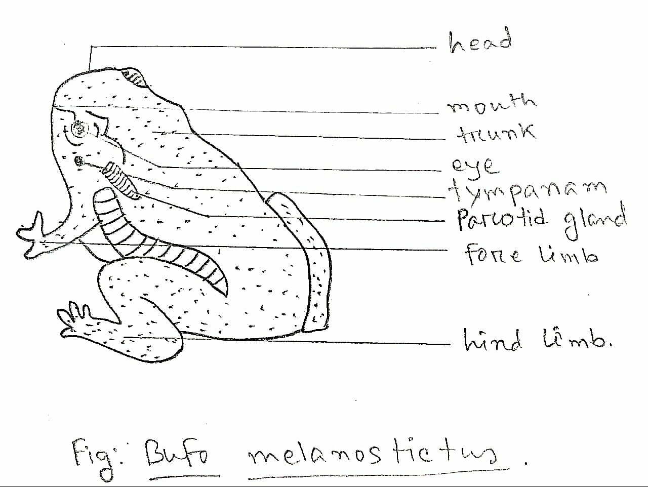
Shape and size
1. Body of a toad is somewhat spindle-shaped, anteriorly pointed, and posteriorly rounded.
2. Body dorso-ventrally flattened.
3. Bilaterally symmetrical body.
4. Bufo melanostictus varies from 57 mm to 83 mm in length.
5. Body is divisible into the head and trunk.
Skin and Color
1. They have thick, dry, naked, and rough skin with prominent cranial ridges and protruding parotid glands.
2. Their skin is also used in respiration and during the hibernation period, the toads respire with their skin.
3. The color pattern is pale yellow-brown; the dorsal side is brownish and the ventral side is yellowish.
4. Toads can change the color of their body to match the surroundings.
5. Postanal tail is absent in the adult stage.
Head
1. The largest transverse aperture, located at the terminal end of the head is the mouth.
2. At the anterior dorsal side of the head and above the mouth, there are two small rounded apertures, known as nostrils. They serve in the respiration of the toad.
3. Two eyes are present on the dorsal side of the head; one on either side of the head.
4. The eyes are very prominent, large, and protruding. Each eye has a thick upper eyelid and a thin ill-developed, semi-transparent lower eyelid.
5. The eyeball of the eye is covered over by a thin, transparent nictitating membrane which is also known as third eyelid.
6. Behind and below each eye, there is a circular area known as the tympanum or eardrum.
7. External ear is absent in toads. The tympanum or eardrum works as the receptor of sound waves.
8. Behind each tympanum, there is a gland on either side of the head called the parotid gland. It secrets poisonous liquid and it works as the defensive organ of toads.
9. In male toads, there are vocal sacs inside the throat. With the help of these vocal sacs, the male toads keep croaking during the breeding season in search of the female.
Trunk
1. Head is joined behind the trunk. The trunk is the broad, short, and flattened part of the body of a toad.
2. Numerous small elevations, known as warts, are present on the dorsal side of the body.
3. There are two pairs of limbs present in the trunk region.
4. Limbs are unequal in size and forelimbs are shorter than the hindlimbs.
5. Forelimbs are consists of brachium, ante-brachium, wrist, and manus(hand). The hand has four digits.
6. In male toads, there is a thumbpad develops during the breeding season which helps the male’s grip during amplexus. Amplexus is a type of mating behavior exhibited by Asian common toads.
7. Hindlimbs are much longer and stronger than forelimbs.
8. Each hindlimb consists of the femur(thigh), crus (shank), and pes (foot). The foot has five elongated digits.
9. The digits of the hindlimbs are jointed and help the toad in swimming.
10. Hindlimbs are modified for jumping and swimming.
11. At the posterior end of the body there is an aperture located between the hindlimbs, is cloaca.
Glands
1. Two types of glands are present on the dorsal side of the toad’s body.
2. The two types of glands are: 1. Mucous glands, 2. Parotid glands or poison glands.
3. There are numerous bumps or warts present on the skin of the dorsal side of the body. These are the mucous glands. Mucous glands are much smaller than the poison glands.
4. Mucous glands keep the skin moist, and facilitate gas exchanges.
5. Parotid glands or poison glands are located behind the tympana and these are the offensive and defensive organs of the toads.
The Nervous System of Asian Common Toad
The nervous system of the toad comprises 1. The central nervous system, 2. The peripheral nervous system, 3. The autonomic nervous system. The nervous system controls and coordinates various activities of the body.
Central Nervous System of Asian Common Toad
1. Central nervous system consists of 1. Brain and 2. Spinal cord.
2. It is a hollow tube whose anterior portion becomes the brain and the posterior portion narrows down to form the spinal cord.
3. The cavity of the brain and the spinal cord is filled up with cerebrospinal fluid.
4. The central nervous system (the brain and the spinal cord) is made up of nerve cells (neurons) and nerve fibers.
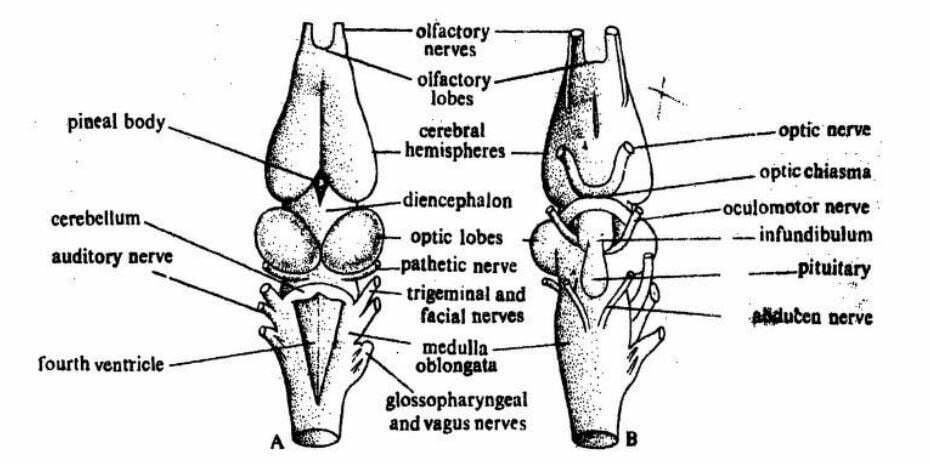
Brain of Asian Common Toad
1. The brain is located inside the cranium.
2. It is differentiated into three parts: forebrain (prosencephalon), midbrain (mesencephalon), and hindbrain (rhombencephalon).
3. Prosencephalon is the anteriormost part of the brain and it is further divided into two parts. Anterior telencephalon and posterior diencephalon.
4. A pair of small olfactory lobes arise from the telencephalon and these lobes are responsible for the sense of smell. The rest of the telencephalon comprises two lobes called the cerebrum or cerebral hemispheres.
5. The roof of the cerebrum is very thin and the floor of the cerebrum is very thick.
6. Behind the cerebrum, the depressed part of the brain is called the diencephalon.
7. The ventral side of the diencephalon bears ‘X’ shaped optic chiasma which is formed by the optic nerves and responsible for the sense of sight.
8. Optic thalami are present on the lateral sides of the diencephalon.
9. The midbrain remains undivided.
10. The optic lobes are present in the midbrain.
11. The posteriormost part of the brain, the rhombencephalon is divided into two parts 1. Metencephalon and 2. Myelencephalon.
12. A thin transverse band like the cerebellum is present in the metencephalon.
13. Medulla oblongata is present in the myelencephalon.
14. The cerebellum coordinates the movement of the body and the medulla oblongata controls and regulates some vital processes like regulation of heartbeat, metabolism, respiration and etc.
15. Some well-formed internal cavities, called ventricles, are present in the brain.
Spinal Cord of Asian Common Toad
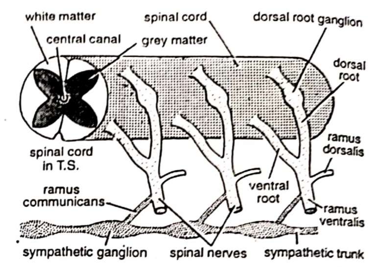
1. The spinal cord is a hollow tube.
2. It extends posteriorly from the medulla oblongata.
3. It remains encased within the neural canal of the vertebral column.
4. The spinal cord has a dorsal fissure and a ventral fissure on its middorsal and midventral lines.
5. The cavity of the spinal cord is known as neurocoel.
Peripheral Nervous System of Asian Common Toad
The peripheral nervous system comprises of the cranial nerves and spinal nerves arising from the cerebrospinal axis. The nerve fibers are of two types and they are 1. afferent or sensory nerve fibers and 2. efferent or motor nerve fibers. The sensory fibers convey the information from the receptor organs to the central nervous system and the motor fibers carry impulses from the central nervous system to the effector organs. Mixed-type nerves are also present which are composed of both sensory and motor nerve fibers.
Cranial Nerves of Asian Common Toad
There are ten pairs of cranial nerves that originate from the brain. Here is the table of the cranial nerves of the Asian common toad.

Spinal Nerves of Asian Common Toad
1. There are ten pairs of spinal nerves present in the toad.
2. All the spinal nerves are mixed types.
3. The first spinal nerve, hypoglossal, supplies to the muscles of the tongue.
4. The second and third spinal nerves of a toad form the brachial plexus.
5. The fourth, fifth, and sixth spinal nerves of a toad supply the integument and the muscles of the trunk region.
6. The seventh, eighth, ninth, and tenth spinal nerves form the sciatic plexus.
Autonomic Nervous System of Asian Common Toad
1. The activities of the autonomic nervous system are somewhat independent.
2. This nervous system regulates the involuntary activities of the body like heartbeat and peristalsis of the alimentary canal.
3. The autonomic nervous system consists of two sympathetic trunks.
4. The sympathetic trunks are located on either side of the dorsal aorta.
5. The sympathetic trunks supply the branches to innervate the cardiac muscles, blood vessels, and alimentary canal.
Sense Organs of Asian Common Toad
The sense organs are the receptors for external stimuli. These organs receive sensations or stimuli due to changes in the external or internal environment. With these sense organs, the central nervous system keeps the information about the outside environment. Like man, toads have five types of special sense organs and they are
1. Sense organ of touch,
2. Sense organ of taste,
3. Sense organ of smell,
4. Sense organ of sight and
5. Sense organ of hearing and balancing.
Sense organs of touch
1. The receptors or organs of touch have another name called tangoreceptors.
2. Toads have numerous microscopic cutaneous sense organs under the epidermis of the skin.
3. These tangoreceptors are sensitive to various types of stimuli such as touch, temperature, humidity, chemicals, light, pain, etc.
Sense organs of taste
1. The receptors or organs of taste have another name called Gustatoreceptors.
2. Taste organs are in the form of taste buds present in small papillae on the tongue and floor and roof of the buccal cavity.
3. Taste buds are made up of two types of cells; the taste cells and the supporting cells.
4. The taste cells are innervated by nerve fibers of facial and glossopharyngeal cranial nerves.
Sense organs of smell
1. The receptors or organs of smell have another name called Olfactoreceptors.
2. These receptors are scattered in the nasal passage.
3. The Olfactoreceptors have two types of cells; olfactory cells and supporting cells.
4. Receptor cells have fine cilia on their outer ends and are attached to the nerve fibers on their inner ends. The fibers unite to form the olfactory nerve which carries the odors to the brain.
5. Supporting cells are simple epithelial cells.
Sense organs of sight
1. The receptors or organs of sight have another name called Photoreceptors.
2. The two large eyes are the photoreceptors of the toads.
3. Each eye has a spherical body called the eyeball.
3. Both eyeballs are spherical in shape and lodged inside an orbit.
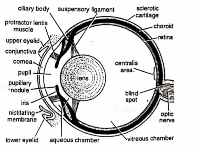
fig: Eyeball of Asian common Toad
4. The eyes can be moved inside the orbit with the help of six extrinsic muscles. The muscles are 1. Superior rectus, 2. Inferior rectus, 3. External rectus, 4. Internal rectus, 5. Superior oblique, and 6. Inferior oblique.
5. Toads can protrude their eyes with the help of levator bulbi and they can withdraw their eyes with the help of retractor bulbi.
6. The eyeball of the toad is made of three concentric layers or coats, an outermost sclerotic, a middle choroid, and an inner retina.
A. Sclerotic Layer
1. The outermost layer of the eyeball is the sclerotic layer.
2. The sclerotic layer is divided into two parts. The anterior part is the cornea and the posterior part is the white of the eye-ball.
3. Cornea is the transparent, circular, and anterior part of the sclerotic layer.
4. Cornea permits the light rays to enter into the eye.
5. Cornea is covered with thin and transparent conjunctiva.
B. Choroid Layer
1. Choroid layer is located underneath the sclerotic layer.
2. Choroid is a richly vascularized and pigmented layer.
3. At the anterior end of the choroid, just behind the cornea, the choroid forms a pigmented circular disc called the iris.
4. The iris has an aperture at the center named pupil.
5. The iris has circular and radial muscles. The pupil can contract and dilate with the help of these muscles.
6. The whole eyeball is lightproof except the pupil.
7. The iris regulates the entry of light into the eyeball.
8. Suspensory ligaments are located just behind the iris and they keep the lens in position.
C. Retinal Layer
1. The innermost layer of the eyeball is the retinal layer.
2. This is the only light-sensitive part of the eyeball.
3. Retina contains two types of photosensitive cells called the rod cells and the cone cells.
4. Cone cells work in the bright light and the vision is colorful.
5. Rod cells work in the low light and the vision is colorless.
6. Photosensitive cells are connected with the optic nerves.
7. The spot where the optic nerve comes out of the eyeball is called the blind spot. It lacks rod and cone cells so it can’t form an image.
8. Light falling on the retina stimulates rods and cones and the impulses are conducted by the optic nerve to the brain.
Mechanism of Sight/Vision
1. Light rays pass through the cornea, pupil, and lens and reach the retina.
2. In the retinal layer, an inverted and reduced image is formed.
3. Inverted image is transmitted to the brain with the help of optic nerves.
4. Then the brain adjusts the image.
5. Toads are myopic and can efficiently see only near objects.
Sense organs of hearing and balancing
1. The two ears are the sense organs of hearing and balancing.
2. The ear of toads consists of two parts and they are middle ear and internal ear.
3. There is no external ear in the toads but some scientists say that the tympanum is the external ear of the toads.
A. Middle Ear
-
-
- The middle ear is a tube-like cavity called the tympanic cavity.
- It is externally closed by the tympanum.
- Middle ear communicates with the pharynx with the Eustachian tube.
- The bony partition between the tympanic cavity and auditory capsule(internal ear) is perforated by an oval aperture called the fenestra ovalis.
- Fenestra ovalis remains closed by a membrane and a cartilaginous nodule, the stapedial plate.
- There is a slender club-like rod of bone and cartilage named columella auris, that extends across the tympanic cavity.
- The outer end of columella auris is attached to the tympanum/eardrum and the inner end is attached to the stapedial plate.
-
B. Internal Ear
-
-
- The membranous labyrinth represents the internal ear whose cavity is filled with milky fluid, called the endolymph.
- The membranous labyrinth remains enclosed in the auditory cavity and floats on the fluid named perilymph.
- The auditory capsule remains closed on all sides by a membranous partition.
- Membranous labyrinth has two chambers. The upper chamber is the utriculus and the lower chamber is the sacculus.
- Three narrow tubular semi-circular canals arise from the utriculus.
- The sacculus produces a short projection called lagena.
- Patches of sensory receptors are present in the inner wall of the membranous labyrinth and each receptor cell is connected with a fiber from the auditory nerve.
-
————– THE END ————–
See more:
- The Nervous System of Frog | Diagram
- Digestive system of Hoplobatrachus tigerinus (Frog) with Diagram – Indian Bull Frog
- External Morphology of Frog | Hoplobatrachus tigerinus | Diagram
- General Characters of All Classes of Vertebrates.
Reference:

Md Ekarm Hossain Bhuiyan is a dedicated zoology graduate with a profound passion for the study of animal life. He completed his primary and secondary education at Ispahani Public School and College, renowned for its commitment to academic excellence. He then pursued his secondary education at Government Science College. After that he achieved graduation at Department of Zoology, Jagannath University. His educational background and enthusiasm for zoology position him to make meaningful contributions to the field of biological sciences in Bangladesh.